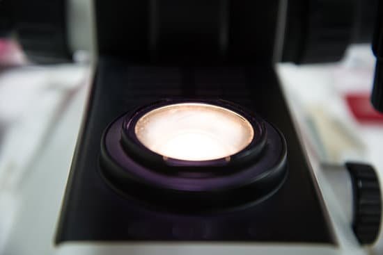
What can you view with a dissecting microscope? A dissecting microscope is used to view three-dimensional objects and larger specimens, with a maximum magnification of 100x. This type of microscope might be used to study external features on an object or to examine structures not easily mounted onto flat slides.
Can you see cells with a dissecting microscope? A dissection microscope is light illuminated. The image that appears is three dimensional. It is used for dissection to get a better look at the larger specimen. You cannot see individual cells because it has a low magnification.
Can the dissecting microscope only view non living things? Both are electron microscopes, but the dissecting microscope has a higher magnification than the compound light microscope. Both are electron microscopes, but the compound light microscope can view only nonliving things.
What objects could be recommended as specimens for viewing under the dissecting microscope? As mentioned, a dissecting (stereo or inspection microscope) is a low power microscope that is commonly used for the purposes of inspecting larger sized specimen/objects like fossils, rocks, insects, and parts of a plant, etc. Here, however, specimen mounted on slides can also be viewed using this type of microscope.
What can you view with a dissecting microscope? – Related Questions
How big is red algae under a microscope?
The SCRP clade are microalgae, consisting of both unicellular forms and multicellular microscopic filaments and blades. The BF are macroalgae, seaweed that usually do not grow to more than about 50 cm in length, but a few species can reach lengths of 2 m.
Who found the light microscope?
The Dutch spectacle maker Hans Janssen and his son Zacharias are generally credited with creating these compound microscopes. The two of them built what was probably the first compound microscope in the last decade of the 16th century.
How does water act as a lens in a microscope?
Due to something called the capillary effect, however, a layer of water in a cup shows a surface that is slightly bent inward. It will act as a concave lens that bends the light rays outward. As a result, letters seen through the layer of water in a cup appear smaller than they are.
Are tardigrades microscopic?
Tardigrades are microscopic eight-legged animals that have been to outer space and would likely survive the apocalypse. … Around 1,300 species of tardigrades are found worldwide.
What does the condenser knob on a microscope do?
Condenser Focusing Knob – This control is used to precisely adjust the vertical height of the condenser. Condenser Lens – This lens system is located immediately under the stage and focuses the light on the specimen.
How do electron microscopes have higher resolution?
As the wavelength of an electron can be up to 100,000 times shorter than that of visible light photons, electron microscopes have a higher resolving power than light microscopes and can reveal the structure of smaller objects.
Who improved the original microscope?
It fell to a Dutch scientist, Anton van Leeuwenhoek, to make further improvements. Van Leeuwenhoek is sometimes popularly credited with the microscope’s invention.
How much can an electron microscope magnify an object?
Transmission electron microscopes (TEM) are microscopes that use a particle beam of electrons to visualize specimens and generate a highly-magnified image. TEMs can magnify objects up to 2 million times. In order to get a better idea of just how small that is, think of how small a cell is.
What features are best for a high school student microscope?
For both college and high school students, it is advisable to go for a microscope with a wide-field eyepiece. These are typically ones with a larger lens opening. Wide-field eyepieces are beneficial for users because it’s easier to position the eyes in order to see into the eyepiece.
Are there microscopes that can see atoms?
The very powerful microscopes are called atomic force microscopes, because they can see things by the forces between atoms. So with an atomic force microscope you can see things as small as a strand of DNA or even individual atoms.
What are the microscopic hairs in your taste buds?
See all those bumps? Those are called papillae (say: puh-PILL-ee), and most of them contain taste buds. Taste buds have very sensitive microscopic hairs called microvilli (say: mye-kro-VILL-eye). Those tiny hairs send messages to the brain about how something tastes, so you know if it’s sweet, sour, bitter, or salty.
How to tell bv or yeast infection through a microscope?
A white, lumpy discharge that looks like cottage cheese may mean a vaginal yeast infection is present. A yellow-green, foamy discharge that has a bad odor may mean trichomoniasis is present. A thin, gray-white vaginal discharge with a strong fishy odor may mean bacterial vaginosis is present.
What is racking of microscope?
Rack and Pinion Focusing Mechanism. A metal rack and pinion used in better quality microscopes for focusing purposes and moving mechanical stages. Rack Stop. A safety feature that prevents the viewer from allowing the objective lens to accidentally hit the stage and damage the specimen or slide.
What do you call a photo from microscope?
A micrograph or photomicrograph is a photograph or digital image taken through a microscope or similar device to show a magnified image of an object. … Micrography is the practice or art of using microscopes to make photographs.
Which microscope to identify asbestos?
There are two different types of electron microscope used for asbestos analysis: Scanning Electron Microscope (SEM) and Transmission Electron Microscope (TEM). Scanning Electron Microscopy is useful in identifying minerals.
How many lenses do microscopes use?
A typical microscope has three or four objective lenses with different magnifications, screwed into a circular “nosepiece” which may be rotated to select the required lens. These lenses are often color coded for easier use. The least powerful lens is called the scanning objective lens, and is typically a 4× objective.
Why would i have microscopic blood in my urine?
Microscopic urinary bleeding is a common symptom of glomerulonephritis, an inflammation of the kidneys’ filtering system. Glomerulonephritis may be part of a systemic disease, such as diabetes, or it can occur on its own.
Who created scanning transmission electron microscope?
Four years later, in 1937, Siemens financed the work of Ernst Ruska and Bodo von Borries, and employed Helmut Ruska, Ernst’s brother, to develop applications for the microscope, especially with biological specimens. Also in 1937, Manfred von Ardenne pioneered the scanning electron microscope.
What is the smallest an electron microscope can see?
Light microscopes let us look at objects as long as a millimetre (10-3 m) and as small as 0.2 micrometres (0.2 thousands of a millimetre or 2 x 10⁻⁷ m), whereas the most powerful electron microscopes allow us to see objects as small as an atom (about one ten-millionth of a millimetre or 1 angstrom or 10-10 m).
What’s the difference between compound and dissecting microscopes?
Dissecting and compound light microscopes are both optical microscopes that use visible light to create an image. … Most importantly, dissecting microscopes are for viewing the surface features of a specimen, whereas compound microscopes are designed to look through a specimen.
When were first microscopes developed?
The first compound microscopes date to 1590, but it was the Dutch Antony Van Leeuwenhoek in the mid-seventeenth century who first used them to make discoveries. When the microscope was first invented, it was a novelty item.

