What cell parts can you see with the microscope? Using a light microscope, one can view cell walls, vacuoles, cytoplasm, chloroplasts, nucleus and cell membrane. Light microscopes use lenses and light to magnify cell parts.
What cell structures are visible with a light microscope? Note: The nucleus, cytoplasm, cell membrane, chloroplasts and cell wall are organelles which can be seen under a light microscope.
What are the parts of the cell visible? The major parts of a cell are the nucleus, cytoplasm, and cell membrane. For plant cells, there is a cell wall. The cytoplasm contains organelles suspended in fluid.
What is the only part of the cell that will be visible under the microscope? Usually, only the cell’s nucleus is visible through a light microscope. In plant cells, the cell wall and vacuole may also be visible. However, there are many cell parts (also called organelles) that are too small to be seen through a light microscope. Three examples of these organelles are…
What cell parts can you see with the microscope? – Related Questions
How do u switch objectives on a microscope?
When focusing on a slide, ALWAYS start with either the 4X or 10X objective. Once you have the object in focus, then switch to the next higher power objective. Re-focus on the image and then switch to the next highest power.
Can you see spindle fibers in plant cells under microscope?
When viewed using a light microscope, the “spindle” (named after a device used for spinning thread) looks like a hairy, elongated ball originating (in animal cells) from the asters around the centrioles, or from opposite sides of the plant cell.
How to find the magnification of light microscope?
To calculate the total magnification of the compound light microscope multiply the magnification power of the ocular lens by the power of the objective lens. For instance, a 10x ocular and a 40x objective would have a 400x total magnification. The highest total magnification for a compound light microscope is 1000x.
When should you use the scanning lens on the microscope?
When should you use the scanning lens on the microscope? Whenever a new slide is viewed or when the view of the specimen in the field of view of a higher-power lens is lost.
How do you carry the compound microscope?
Always keep your microscope covered when not in use. Always carry a microscope with both hands. Grasp the arm with one hand and place the other hand under the base for support.
How do i get more magnification from my microscope?
If you change your standard 10X eyepieces, you can increase your overall magnification without changing your working distance ( the space between the lens and the specimen). By adding an auxiliary lens, you can either increase or decrease magnification however the working distance will change.
What is the range of magnification of the microscope?
Objectives typically have magnifying powers that range from 1:1 (1X) to 100:1 (100X), with the most common powers being 4X (or 5X), 10X, 20X, 40X (or 50X), and 100X.
What to look for in a good microscope?
When buying a compound microscope, always ensure that the microscope has an iris diaphragm and good quality condenser – ideally, an Abbe condenser which allows for greater adjustments. Both items are found in the sub-stage of the microscope and are used in adjusting the base illumination.
What is the nosepiece of a microscope called?
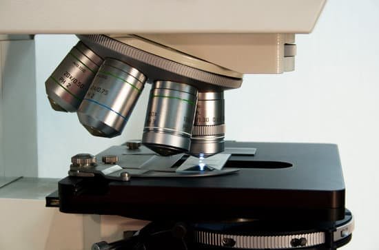
Nosepiece: The upper part of a compound microscope that holds the objective lens. Also called a revolving nosepiece or turret.
What do eosinophils look like under a microscope?
Eosinophils contain large granules, and the nucleus exists as two nonsegmented lobes. In addition, the granules of eosinophils typically stain red, which makes them easily distinguished from other granulocytes when viewed on prepared slides under a microscope.
Are scabies mites microscopic?
hominis). The microscopic scabies mite burrows into the upper layer of the skin where it lives and lays its eggs. The most common symptoms of scabies are intense itching and a pimple-like skin rash. The scabies mite usually is spread by direct, prolonged, skin-to-skin contact with a person who has scabies.
What is type c7 in microscope?
Type:C7. Features. -100% Brand new and high quality. -0.1 coordinate eyepiece scale, vertical and horizontal coordinates of the length of 10mm, divided into 100 equal parts, each index value of 0.1mm, slide diameter Φ19mm. -Mainly used for correction of microscopes and other optical devices.
What does a eyepiece do on a microscope?
The eyepiece, or ocular lens, is the part of the microscope that magnifies the image produced by the microscope’s objective so that it can be seen by the human eye.
Why is a convex lens used in a microscope?
For the purposes of microscopy, convex lenses are used for their ability to focus light at a single point. … Microscopes borrowed this idea, using convex lenses to focus light towards a point that is f distance away from the lens. This distance is known as the focal length of the lens and depends on the shape.
Can microscopic organisms be sentient?
Beings that have no centralized nervous systems are not sentient. This includes bacteria, archaea, protists, fungi, plants and certain animals.
What magnifies the image 400 times on a microscope?
The total magnification of a high-power objective lens combined with a 10x eyepiece is equal to 400x magnification, giving you a very detailed picture of the specimen in your slide.
Does a compound microscope use real or virtual image?
With the compound microscope, this intermediate image is real, formed by the objective lens. In all cases, the function of the eyepiece is to form a virtual, magnified image for your eye to view.
Can we see atoms under a microscope?
Atoms are really small. So small, in fact, that it’s impossible to see one with the naked eye, even with the most powerful of microscopes. … Now, a photograph shows a single atom floating in an electric field, and it’s large enough to see without any kind of microscope.
Which part of the microscope are used to carry it?
The three basic, structural components of a compound microscope are the head, base and arm. Arm connects to the base and supports the microscope head. It is also used to carry the microscope.
Why scientist use microscope?
Scientists use microscopes to observe objects too small to view with the human eye. Microscopes can magnify an image hundreds of times while…
What does strep look like under microscope?
Under a microscope, streptococcus bacteria look like a twisted bunch of round berries. Illnesses caused by streptococcus include strep throat, strep pneumonia, scarlet fever, rheumatic fever (and rheumatic heart valve damage), glomerulonephritis, the skin disorder erysipelas, and PANDAS. Familiarly known as strep.

