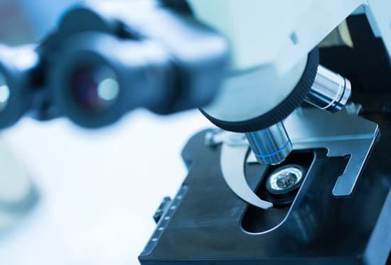
What country was the microscope invented in? The microscope was invented by the Dutch spectacle maker Zaccharias Janssen around 1590. This was the time when Toyotomi Hideyoshi was unifying Japan into a single nation. In 1655, the Englishman Robert Hooke produced a “compound microscope” that included an objective lens and an eyepiece lens.
Who invented the microscope and where? The development of the microscope allowed scientists to make new insights into the body and disease. It’s not clear who invented the first microscope, but the Dutch spectacle maker Zacharias Janssen (b. 1585) is credited with making one of the earliest compound microscopes (ones that used two lenses) around 1600.
Was the microscope invented in Italy? The Campani compound microscope is a microscope on exhibit at the Museo Galileo in Italy, thought to have been built by optical instrument maker Giuseppe Campani in the second half 17th century.
Who made first microscope? A Dutch father-son team named Hans and Zacharias Janssen invented the first so-called compound microscope in the late 16th century when they discovered that, if they put a lens at the top and bottom of a tube and looked through it, objects on the other end became magnified.
What country was the microscope invented in? – Related Questions
Is nucleus visible under light microscope?
Note: The nucleus, cytoplasm, cell membrane, chloroplasts and cell wall are organelles which can be seen under a light microscope.
What are some examples of microscopic organisms?
A microorganism is a living thing that is too small to be seen with the naked eye. Examples of microorganisms include bacteria, archaea, algae, protozoa, and microscopic animals such as the dust mite.
How do the two types of electron microscopes work?
Here we compare two basic types of microscopes – optical and electron microscopes. The electron microscope uses a beam of electrons and their wave-like characteristics to magnify an object’s image, unlike the optical microscope that uses visible light to magnify images.
How do microscopes help scientists today?
A microscope is an instrument that is used to magnify small objects. Some microscopes can even be used to observe an object at the cellular level, allowing scientists to see the shape of a cell, its nucleus, mitochondria, and other organelles.
What is the position of the image under the microscope?
The optics of a microscope’s lenses change the orientation of the image that the user sees. A specimen that is right-side up and facing right on the microscope slide will appear upside-down and facing left when viewed through a microscope, and vice versa.
What is the best microscope to view coins?
For coins, a good range of magnification is 10x-30x. The image at right shows a penny at 20x magnification.
Why must specimens viewed with a compound microscope be thin?
A specimen has to be thin so that the light coming from the light source is able to pass through the specimen Specimens are sometimes stained with dyes so that they are easier to distinguish and find.
How has the microscope revolutionized medicine?
The microscope has had a major impact in the medical field. Doctors use microscopes to spot abnormal cells and to identify the different types of cells. This helps in identifying and treating diseases such as sickle cell caused by abnormal cells that have a sickle like shape.
Why does each microscope need to be calibrated independently?
You usually get a more accurate estimate of cell size when viewing them at a higher magnification. This procedure describes how to calibrate your microscope to estimate cell size. As every microscope is a bit different, each microscope should be calibrated independently.
What is the meaning of microscope stage?
Microscope Stages. All microscopes are designed to include a stage where the specimen (usually mounted onto a glass slide) is placed for observation. Stages are often equipped with a mechanical device that holds the specimen slide in place and can smoothly translate the slide back and forth as well as from side to side …
What are the three lenses on microscope?
Compound microscopes use three lenses are used to accomplish the viewing, the eyepiece lens, condenser lens and objective lens. There can also be compound and electron lenses and these lend themselves to the power and magnification of the device itself.
What did robert koch discover by using a compound microscope?
In the medical world, Louis Pasteur of France used a compound microscope to discover yeast fungus, while Karl J. Ebert, a German bacteriologist, employed a compound microscope in his discovery of Eberthella Thyphosa. It was also a compound microscope that Robert Koch discover tubercle and cholera bacilli.
How to write microscope magnification?
100X (this means that the image being viewed will appear to be 100 times its actual size).
How many types of microscope we have?
There are several different types of microscopes used in light microscopy, and the four most popular types are Compound, Stereo, Digital and the Pocket or handheld microscopes. Some types are best suited for biological applications, where others are best for classroom or personal hobby use.
How do the optical parts of a microscope achieve magnification?
In simple magnification, light from an object passes through a biconvex lens and is bent (refracted) towards your eye. … Both of these contribute to the magnification of the object. The eyepiece lens usually magnifies 10x, and a typical objective lens magnifies 40x.
What is microscopic polyarteritis nodosa?
Recently individualized from PAN, microscopic polyangiitis (MPA) is a systemic vasculitis of small-size vessels whose clinical manifestations are very similar to those of PAN, but it is characterized by the presence of rapidly progressive glomerulonephritis (RPGN), which is nearly constant, and pulmonary involvement …
How to see sperm cell in microscope?
You can view sperm at 400x magnification. You do NOT want a microscope that advertises anything above 1000x, it is just empty magnification and is unnecessary. In order to examine semen with the microscope you will need depression slides, cover slips, and a biological microscope.
What are the microscopic differences between synthetic and natural gems?
Synthetic gemstones, while created in a lab, have exactly the same chemical composition as the natural one. For instance, a diamond is made out of carbon. The only difference between a natural diamond and a lab-created diamond is how that carbon was compressed into a gem, either by nature or by technology.
How much does an optical microscope cost?
A typical optical microscope might cost $20,000 to $40,000, whereas an electron microscope can cost several million dollars, he said.
How is a brightfield microscope used?
Brightfield Microscope is used in several fields, from basic biology to understanding cell structures in cell Biology, Microbiology, Bacteriology to visualizing parasitic organisms in Parasitology. Most of the specimens to viewed are stained using special staining to enable visualization.
How much does a light microscope magnify?
Light microscopes allow for magnification of an object approximately up to 400-1000 times depending on whether the high power or oil immersion objective is used. Light microscopes use visible light which passes and bends through the lens system.

