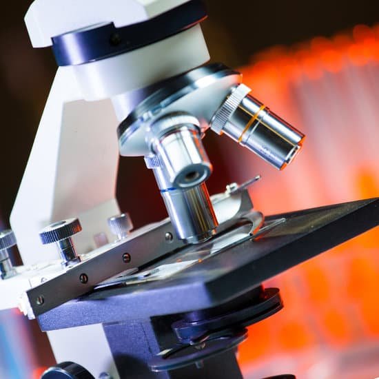
What did the first people who used microscopes look at? Enter Anton van Leeuwenhoek, who used a microscope with one lens to observe insects and other specimen. Leeuwenhoek was the first to observe bacteria.
What did people first look at when microscopes were invented? The early simple “microscopes” which were really only magnifying glasses had one power, usually about 6X – 10X . One thing that was very common and interesting to look at was fleas and other tiny insects. These early magnifiers were hence called “flea glasses”.
What was the microscope first used for? Grinding glass to use for spectacles and magnifying glasses was commonplace during the 13th century. In the late 16th century several Dutch lens makers designed devices that magnified objects, but in 1609 Galileo Galilei perfected the first device known as a microscope.
Who was the first to use a microscope to look at living things? Using his microscope, Leeuwenhoek was the first person to observe human cells and bacteria. Figure 5.2. 2: Robert Hooke sketched these cork cells as they appeared under a simple light microscope.
What did the first people who used microscopes look at? – Related Questions
Can atoms be seen under a regular microscope?
Atoms are so small that it’s almost impossible to see them without microscopes. … The diameter of a strontium atom is a few millionths of a millimeter.
Why must specimens with a compound microscope be thin?
A specimen has to be thin so that the light coming from the light source is able to pass through the specimen Specimens are sometimes stained with dyes so that they are easier to distinguish and find.
How to align microscope?
To align, turn the brightness knob down to a fairly low setting, then remove the frosted glass filter from the light path. On an upright microscope place a piece of lens paper over the field diaphragm to see the image of the filament.
Which oil is used in oil immersion microscope?
Only use oil which is recommended by the objective manufacturer. For many years, cedar wood oil was routinely used for immersion (and is still commercially available). Although this oil has a refractive index of 1.516, it has a tendency to harden and can cause lens damage if not removed after use.
Why replace toothbrush microscope?
Krasnow found that “after just a few months the toothbrush is smooth, worn down by the abrasive effects of the toothpaste, rendering it nearly ineffective for proper dental hygiene.” Medical Daily says that “under a microscope, a three-month old brush looks pretty useless.” According to the ADA, “consumers should …
How to compute total magnification in microscope?
To figure the total magnification of an image that you are viewing through the microscope is really quite simple. To get the total magnification take the power of the objective (4X, 10X, 40x) and multiply by the power of the eyepiece, usually 10X.
How does a compound microscope magnify images?
It is through the microscope’s lenses that the image of an object can be magnified and observed in detail. … When light reflects off of an object being viewed under the microscope and passes through the lens, it bends towards the eye. This makes the object look bigger than it actually is.
What is the principle of transmission electron microscope?
The mechanism of a light microscope is that an increase in resolution power decreases the wavelength of the light, but in the TEM, when the electron illuminates the specimen, the resolution power increases increasing the wavelength of the electron transmission.
What are monocular microscopes used for?
Monocular microscopes, microscopes that are equipped with one eye piece, can magnify samples up to 1,000 times. If you need a microscope that magnifies at higher levels, a binocular microscope is right for you. Monocular microscopes are often used in classrooms and laboratories for observing slide samples.
When to use the condenser of a microscope?
On upright microscopes, the condenser is located beneath the stage and serves to gather wavefronts from the microscope light source and concentrate them into a cone of light that illuminates the specimen with uniform intensity over the entire viewfield.
Do nature microscope?
Kenko Do Nature portable microscope is a lightweight handheld microscope, very convenient and can be carried with you everywhere. With a 60 to 120x zoom power, you can change the magnifying power according to the object you would like to see.
What organelles can you not see with a light microscope?
Some cell parts, including ribosomes, the endoplasmic reticulum, lysosomes, centrioles, and Golgi bodies, cannot be seen with light microscopes because these microscopes cannot achieve a magnification high enough to see these relatively tiny organelles.
What is the purpose of the microscope?
A microscope is an instrument that can be used to observe small objects, even cells. The image of an object is magnified through at least one lens in the microscope. This lens bends light toward the eye and makes an object appear larger than it actually is.
What are 2 procedures to handle a light microscope?
When carrying the light microscope, handlers must put one hand on the base at all times, to avoid dropping it, while the other hand should be on the arm. The microscope must never be carried upside down, since the ocular will fall out. It should never be swung when it is carried, according to Miami University.
Who discovered microbes in pond water by using a microscope?
Antonie van Leeuwenhoek used single-lens microscopes, which he made, to make the first observations of bacteria and protozoa.
Cuál es la unidad de medida utilizada en microscopía?
¿Cuáles son las unidades de medida del microscopio? Un micrómetro, también conocido como micra/micrón, es una unidad de medida microscópica equivalente a 0,001 mm o 0,000039 pulgadas.
How did the microscope help understanding cells?
Microscopes allow humans to see cells that are too tiny to see with the naked eye. … These cells are small and contain no membrane- bound organelles. It allowed them to observe Eukaryotic cells with a nucleus and membrane-bound organelles that perform different life functions.
Can you see spirochetes under microscope?
Many spirochetes are difficult to see by routine microscopy. Although they are gram negative, many either take stains poorly or are too thin (0.15 μm or less) to fall within the resolving power of the light microscope.
Which microscope can be used to visualize non living things?
Electron microscopy (EM) is a technique for obtaining high resolution images of biological and non-biological specimens. It is used in biomedical research to investigate the detailed structure of tissues, cells, organelles and macromolecular complexes.
What does microscopic exam stand for?
This test looks at a sample of your urine under a microscope. It can see cells from your urinary tract, blood cells, crystals, bacteria, parasites, and cells from tumors. This test is often used to confirm the findings of other tests or add information to a diagnosis.
Which organelles are visible in the light microscope?
Note: The nucleus, cytoplasm, cell membrane, chloroplasts and cell wall are organelles which can be seen under a light microscope.

