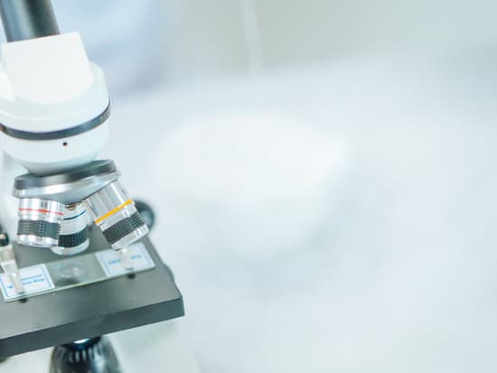
What does inverted mean in microscopes? An inverted microscope is a microscope in which the light source is pointing down onto the stage while the sample is viewed from below.
What does it mean when an image is inverted in microscope? There are also mirrors in the microscope, which cause images to appear upside down and backwards. … The letter appears upside down and backwards because of two sets of mirrors in the microscope. This means that the slide must be moved in the opposite direction that you want the image to move.
What is inversion in a microscope? Inversion is the reversal of an image projected by a microscope. Most microscopes used today are compound microscopes, meaning they have more that one lens involved in the magnification process. … The objective lens is curved, thus causing the light that passes through it to cross over, thus inverting the image.
Why is a microscope inverted? Inverted microscopes are useful for observing living cells or organisms at the bottom of a large container (e.g., a tissue culture flask) under more natural conditions than on a glass slide, as is the case with a conventional microscope.
What does inverted mean in microscopes? – Related Questions
Who made the first stereo microscope?
In the early 1890’s, an American biologist and instrument maker, Horatio S. Greenough developed a stereo microscope which was an alternative design to the CMO microscope.
How does the image through a dissecting microscope move?
A specimen that is right-side up and facing right on the microscope slide will appear upside-down and facing left when viewed through a microscope, and vice versa. Similarly, if the slide is moved left while looking through the microscope, it will appear to move right, and if moved down, it will seem to move up.
How many microscopes did leeuwenhoek make?
Antonie van Leeuwenhoek made more than 500 optical lenses. He also created at least 25 single-lens microscopes, of differing types, of which only nine have survived. These microscopes were made of silver or copper frames, holding hand-made lenses. Those that have survived are capable of magnification up to 275 times.
How hard is it to see sperm under a microscope?
You can view sperm at 400x magnification. You do NOT want a microscope that advertises anything above 1000x, it is just empty magnification and is unnecessary. In order to examine semen with the microscope you will need depression slides, cover slips, and a biological microscope.
Where is the rheostat on a microscope?
The microscope rheostat control can be found on the side of the compound microscope body. It will typically be a knob that is turned clockwise in order to increase the light intensity, or counter-clockwise to reduce the light.
How to calculate magnification of light microscope?
To calculate the total magnification of the compound light microscope multiply the magnification power of the ocular lens by the power of the objective lens. For instance, a 10x ocular and a 40x objective would have a 400x total magnification. The highest total magnification for a compound light microscope is 1000x.
What microscope did robert hooke use to discover cells?
Robert Hooke’s Microscope. Robert Hook refined the design of the compound microscope around 1665 and published a book titled Micrographia which illustrated his findings using the instrument.
What factor limits the resolution barrier of light microscopes?
The resolution for optical microscopy is limited by the diffraction, or the “spreading out,” of the light wave when it passes through a small aperture or is focused to a tiny spot.
Is a refractometer a microscope?
Abbe (Laboratory) Refractometer Abbe Refractometers are bench-top meters that look similar to a microscope which provide highly precise measurements of refractive index.
Why lycopodium powder is used in travelling microscope experiment?
Travelling microscope is used to measure the real and apparent depth of the spot. Complete step-by-step answer: … But a microscope would not focus here since glass is transparent. So lycopodium powder is added on the surface so that we have some reference to focus on.
What is condenser microscope?
On upright microscopes, the condenser is located beneath the stage and serves to gather wavefronts from the microscope light source and concentrate them into a cone of light that illuminates the specimen with uniform intensity over the entire viewfield.
Are there microscopic bugs on eyelashes?
Eyelash mites are microscopic organisms that naturally live at the base of your eyelashes. These tiny eight-legged eyelash bugs actually live in your hair follicles, and feed on dead skin cells and the oils in your skin. Eyelash mites are also known as demodex mites.
When would you use a compound microscope?
Typically, a compound microscope is used for viewing samples at high magnification (40 – 1000x), which is achieved by the combined effect of two sets of lenses: the ocular lens (in the eyepiece) and the objective lenses (close to the sample).
What is metallographic microscope?
Metallographic microscopes are used to identify defects in metal surfaces, to determine the crystal grain boundaries in metal alloys, and to study rocks and minerals. This type of microscope employs vertical illumination, in which the light source is inserted into the microscope tube…
Who named the first microscope?
Lens Crafters Circa 1590: Invention of the Microscope. Every major field of science has benefited from the use of some form of microscope, an invention that dates back to the late 16th century and a modest Dutch eyeglass maker named Zacharias Janssen.
What does the prefix stereo mean in stereo microscope?
before vowels stere-, word-forming element meaning “solid, firm; three-dimensional; stereophonic,” from Greek stereos “solid” (from PIE root *ster- (1) “stiff”).
What is the light adjustment knob on a microscope?
It controls how far the light condenser is from the slide, which should be properly adjusted before you use the microscope. If you move it, you will have it in the wrong position. If your scope has the knob, find out where it is and avoid it.
What is scanning objective on a microscope?
A scanning objective lens provides the lowest magnification power of all objective lenses. … The name “scanning” objective lens comes from the fact that they provide observers with about enough magnification for a good overview of the slide, essentially a “scan” of the slide.
How fast can microscope cameras capture images?
Thanks to a global shutter and the latest CMOS sensor technology, the camera balances high frames rates with resolution. See images in 4K resolution at 32 frames per second (fps) or full HD live images up to 64 fps (the maximum framerate a standard monitor can display).
Why does oil help resolution of microscope?
Key takeaways. The microscope immersion oil decreases the light refraction, allowing more light to pass through your specimen to the objectives lens. Therefore, the microscope immersion oil increases the resolution and improve the image quality.
What would a compound microscope look at examples?
Compound microscopes are used to view small samples that can not be identified with the naked eye. These samples are typically placed on a slide under the microscope. When using a stereo microscope, there is more room under the microscope for larger samples such as rocks or flowers and slides are not required.

