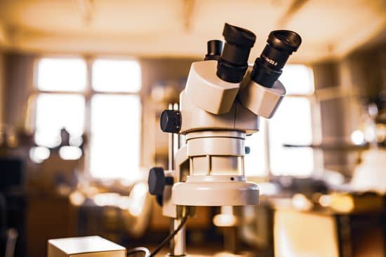
What does microscope mean in latin? First used in the 1650s, microscope is descended from the Modern Latin microscopium, meaning “an instrument for viewing what is small.” In science, microscopes are essential for examining material that can’t be seen with the naked eye, like bacteria and viruses.
What is the meaning of the root word microscope? An easy way to remember that the prefix micro- means “small” is through the word microscope, an instrument which allows the viewer to see “small” living things.
What are the two Latin word of the microscope? The word “microscope” comes from the Latin “microscopium,” which is derived from the Greek words “mikros,” meaning “small,” and “skopein,” meaning “to look at.”
What is Amicroscop? A microscope is a piece of scientific equipment that lets us see very small things that our eyes otherwise couldn’t see. The microscope uses lenses, similar to those in glasses or a magnifying glass, to make things look bigger – this is called magnification. Magnification.
What does microscope mean in latin? – Related Questions
Can the mitochondria be seen by a light microscope?
Mitochondria are visible with the light microscope but can’t be seen in detail. Ribosomes are only visible with the electron microscope.
How to increase contrast on a light microscope?
Contrast may be improved by placing suitable apertures or filters within the optical path, either in the illuminating system alone (dark ground or Rheinberg illumination), or in conjugate planes in the imaging system (e.g. for phase contrast, differential interference contrast or polarised light microscopy).
What kind of lenses are in a microscope?
Microscopes use convex lenses in order to focus light. Image from http://clubsciencekrl.blogspot.com/. Microscope objectives contain lenses but are not as simple as the lenses seen in Fig. 2, making them complex lenses (Fig.
What to use to clean microscope lens?
Dip a lens wipe or cotton swab into distilled water and shake off any excess liquid. Then, wipe the lens using the spiral motion. This should remove all water-soluble dirt.
Who invented microscope?
The development of the microscope allowed scientists to make new insights into the body and disease. It’s not clear who invented the first microscope, but the Dutch spectacle maker Zacharias Janssen (b. 1585) is credited with making one of the earliest compound microscopes (ones that used two lenses) around 1600.
Why are different microscopes used in material science?
Microscopic techniques make it possible to assess the morphology, composition, physical properties, and dynamic behavior of materials, thus making a significant contribution to the development of material science. They are necessary for both the quality control of products and the development of new materials.
What does microscopic field mean?
Definitions of microscopic field. the areas that is visible through a microscope. type of: field, field of view. the area that is visible (as through an optical instrument)
Why is a microscope a valuable tool for scientific research?
Microscopes help the scientists to study the microorganisms, the cells, the crystalline structures, and the molecular structures, They are one of the most important diagnostic tools when the doctors examine the tissue samples.
How do you adjust the resolution on a microscope?
The resolution of a specimen viewed through a microscope can be increased by changing the objective lens. The objective lenses are the lenses that protrude downward over the specimen. Grasp the nose piece. The nose piece is the platform on the microscope to which the three or four objective lenses are attached.
What are the 2 different types of microscopes?
There are several different types of microscopes used in light microscopy, and the four most popular types are Compound, Stereo, Digital and the Pocket or handheld microscopes.
How to test your sperm under a microscope?
To do a home test, a man would have to wait for around five minutes after ejaculation for the semen to liquefy, then apply a small amount to a plastic sheet and press it against the microscope for inspection. This can be done without getting semen on to the phone, says Kobori.
Can a hernia cause microscopic blood in urine?
The presence of urological symptoms and signs such as hematuria, flank pain and hydroureteronephrosis may be seen with an inguinal hernia, but they generally occur when there is associated bladder herniation [4-6].
What type of radiation do electron microscopes emit?
X-rays are produced in the electron microscope whenever the primary electron beam or back scattered electrons strike metal parts with sufficient energy to excite continuous and/or characteristic X-radiation.
What is the definition of stage on a microscope?
Microscope Stages. All microscopes are designed to include a stage where the specimen (usually mounted onto a glass slide) is placed for observation. Stages are often equipped with a mechanical device that holds the specimen slide in place and can smoothly translate the slide back and forth as well as from side to side …
How to use a polarizer light microscope?
Rotate the 10x objective lens into position on the nosepiece. If necessary, push the analyser completely into place so that it is aligned in the light path. Before placing the specimen on the stage, gradually rotate the polariser until the field of view becomes as dark as possible (extinction).
How do microscopes work optics?
The optical or light microscope uses visible light transmitted through, refracted around, or reflected from a specimen. … Some of the lenses in a microscope bend these light waves into parallel paths, magnify and focus the light at the ocular.
How can you increase the resolution of your microscope?
The resolution of a specimen viewed through a microscope can be increased by changing the objective lens. The objective lenses are the lenses that protrude downward over the specimen.
What microscope uses dyes and uv light?
High-Intensity Light, Dyes and Stains. The fluorescence microscope is the most used microscope in the medical and biological fields. These types of microscopes use high-powered light waves to provide unique image viewing options that are unavailable with traditional microscopes.
Do all protists are unicellular microscopic forms of life?
Most protists are microscopic and unicellular, but some true multicellular forms exist. A few protists live as colonies that behave in some ways as a group of free-living cells and in other ways as a multicellular organism.
Does united scope llc make amscope and omax microscopes?
In addition to AmScope, the United Scope family of brands includes these other great products: The OMAX brand is our line of microscopes targeted to professionals.
How to look at blood cells under microscope?
Place a drop of blood onto a microscope slide. Add a drop of stain to the blood to make the cells easier to see. Carefully place a coverslip over the drop of blood. Sliding it slightly along the microscope slide will spread out the blood cells making them easier to see.

