What does monocular microscope mean? Monocular Microscope: A compound microscope with a single eyepiece. Nosepiece: The upper part of a compound microscope that holds the objective lens. Also called a revolving nosepiece or turret.
What is the monocular microscope? Monocular microscopes, microscopes that are equipped with one eye piece, can magnify samples up to 1,000 times. If you need a microscope that magnifies at higher levels, a binocular microscope is right for you. Monocular microscopes are often used in classrooms and laboratories for observing slide samples.
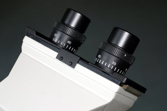
Why is your microscope called a monocular microscope? holds the eyepieces in place above the objective lens. Binocular microscope heads typically incorporate a diopter adjustment ring that allows for the possible inconsistencies of our eyesight in one or both eyes. The monocular (single eye usage) microscope does not need a diopter.
What is the difference between a monocular and a binocular? So, what are the key differences between a Binocular and a Monocular? … For a monocular, it has only one lens that you can hold up to one eye (you can choose to use your left or right eye based on your personal preference), while a binocular comes with 2 lens which you can hold up to both eyes.
What does monocular microscope mean? – Related Questions
Can you see tardigrades without a microscope?
Tardigrades are nearly translucent and they average about half a millimeter (500 micrometers) in length, about the size of the period at the end of this sentence. In the right light you can actually see them with the naked eye.
What does chloroplast look like under a microscope?
Observation – When viewed under the microscope, students will be able to distinguish different parts of the cell including the plastids (chloroplast and mitochondria). On the other hand, a simply wet mount (even without staining) will show chloroplast to be small green (or dark green) sports across the cell surface.
What is another name for light microscope?
The optical microscope, also referred to as a light microscope, is a type of microscope that commonly uses visible light and a system of lenses to generate magnified images of small objects.
How do you get rid of microscopic colitis?
Microscopic colitis can get better on its own, but most patients have recurrent symptoms. The main treatment for microscopic colitis is medication. In many cases, the doctor will start treatment with an antidiarrheal medication such as Pepto-Bismol® or Imodium® .
What kinds of microscopes are used in schools?
The most common types of microscopes used in teaching are monocular light microscopes (80%), followed by binocular optical microscopes (16%), digital microscopes (3%), and stereomicroscopes (1%). A total of 43% of teachers perform microscopy using the demonstration method, and 37% of teachers use practical work.
What muscle types appear striated under a microscope?
Because it can be controlled by thought, skeletal muscle is also called voluntary muscle. Skeletal muscles are long and cylindrical in appearance; when viewed under a microscope, skeletal muscle tissue has a striped or striated appearance.
What is the scanning lens for in a microscope?
A scanning objective lens provides the lowest magnification power of all objective lenses. 4x is a common magnification for scanning objectives and, when combined with the magnification power of a 10x eyepiece lens, a 4x scanning objective lens gives a total magnification of 40x.
What is the parts and function of microscope?
Body tube (Head): The body tube connects the eyepiece to the objective lenses. Arm: The arm connects the body tube to the base of the microscope. Coarse adjustment: Brings the specimen into general focus. Fine adjustment: Fine tunes the focus and increases the detail of the specimen.
How to look at ringworm under microscope?
To be certain of a diagnosis of ringworm, it is imperative to microscopically examine and positively identify the fungus.
Are microvilli microscopic?
Microvilli (singular: microvillus) are microscopic cellular membrane protrusions that increase the surface area for diffusion and minimize any increase in volume, and are involved in a wide variety of functions, including absorption, secretion, cellular adhesion, and mechanotransduction.
Which lens is found within the eyepiece of the microscope?
The image magnified by the objective lens is further magnified by the ocular lens for observation. An ocular lens consists of one to three lenses and is also provided with a mechanism, called a field stop, that removes unnecessary reflected light and aberration.
How did the letter e appear under the microscope?
The letter “e” appears upside down and backwards under a microscope. Either, diatoms are single celled, or they do not have a cell wall.
What is the ocular lens of a microscope?
The ocular lenses are the lenses closest to the eye and usually have a 10x magnification. Since light microscopes use binocular lenses there is a lens for each eye.
Do i dampen the microscope optical lens wipe before using?
Be careful when cleaning the eyepieces as these solvents may damage the rubber eyeshields. Wrap lens tissue over applicator stick or Q-tip as above, and slightly dampen with solvent before wiping.
What kind of microscope should i buy?
You will need a compound microscope if you are viewing “smaller” specimens such as blood samples, bacteria, pond scum, water organisms, etc. … Typically, a compound microscope has 3-5 objective lenses that range from 4x-100x. Assuming 10x eyepieces and 100x objective, the total magnification would be 1,000 times.
What is the difference between optical microscope and electron microscope?
Optical microscopes use a simple lens, whereas electron microscopes use an electrostatic or electromagnetic lens. … Optical microscopes use photons or light energy, while electron microscopes use electrons, which have shorter wavelengths that allows greater magnification.
What a scanning electron microscope is used for?
scanning electron microscope (SEM), type of electron microscope, designed for directly studying the surfaces of solid objects, that utilizes a beam of focused electrons of relatively low energy as an electron probe that is scanned in a regular manner over the specimen.
How does the stereo microscope work?
How Do Stereo Microscopes Work? A stereo or a dissecting microscope uses reflected light from the object. It magnifies at a low power hence ideal for amplifying opaque objects. Since it uses light that naturally reflects from the specimen, it is helpful to examine solid or thick samples.
Can advil cause microscopic blood in urine?
Yes, ibuprofen can cause hematuria (blood in the urine). Due to you having blood in your urine it would most likely be recommended that you do not take ibuprofen or other NSAID in the future, unless you have been prescribed them. Other non-steroidal anti-inflammatory (NSAID) drugs may cause the same side effect.
What’s the difference between a biological microscope and stereoscope?
The Differences Between Biological Microscopes and Stereo Microscopes. … The magnification of a biological microscope is much higher than that of a stereo microscope. Magnification is changed by revolving the nose piece so a new objective lens (either 4x, 10x, 40x or 100x) is in place.
Is a microscopic planktonic algae alive?
Planktonic algae are microscopic plants that live in every drop of pond water. These primitive creatures are extremely important to the aquatic ecosystem because they are the base for the food chain and are largely responsible for the chemistry of the pond.

