What does quartz crystal look like under a microscope? Under the microscope, quartz lacks cleavage and colour and has low first-order, grey-white interference colours (Figure 53b and c). Being chemically stable, quartz crystals look clean compared with feldspars, which are almost always turbid or cloudy.
How do you identify a quartz crystal? Clear quartz may have small inclusions that look like a smudge in the crystal, but overall the crystal should be colorless and transparent. Inspect the crystal shape. Quartz crystals are generally hexagonal prisms that terminate on each end with a six-sided pyramid. … Quartz will streak either white or colorless.
What does real quartz crystal look like? Test the unknown stone under inspection by trying to scratch a common piece of glass such as a glass bottle. If the object easily scratches the glass, the specimen probably is quartz crystal. If scratching the glass takes a lot of effort, the specimen likely is another piece of glass.
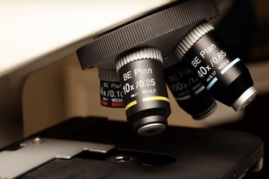
What type of microscope is a light microscope? The common light microscope used in the laboratory is called a compound microscope because it contains two types of lenses that function to magnify an object. The lens closest to the eye is called the ocular, while the lens closest to the object is called the objective.
What does quartz crystal look like under a microscope? – Related Questions
What is the function of an eyepiece on a microscope?
The eyepiece, or ocular lens, is the part of the microscope that magnifies the image produced by the microscope’s objective so that it can be seen by the human eye.
What microscope achieves the highest magnification?
Out of all types of microscopes, the electron microscope has the greatest capability in achieving high magnification and resolution levels, enabling us to look at things right down to each individual atom.
How to use pocket microscope?
Place the microscope directly on the top of the object of observation and press the LED On/Off Button. 2. Slide the Zoom Lever to adjust the desired degree of magnification (from 20x to 40x) 3. Turn the Focusing wheel gradually until the image is clear and sharp.
What does an euglena look like under a microscope?
When viewed under the light microscope, Euglena appear as elongated unicellular organisms that are rapidly moving across the field surface. … Although one flagellum is often seen, they have two flagella, one of which is often hidden in a part of the Euglena referred to as reservoir.
What is microscopic agglutination test?
INDIRECT READING. MAT is a serologic technique that detects agglutinating antibodies against serovar- associated epitopes of the microorganism Leptospira spp. Equal volumes of serial diluted sera are mixed with live leptospires.
Are there microscopic bugs on your skin?
It might give you the creepy-crawlies, but you almost certainly have tiny mites living in the pores of your face right now. They’re known as Demodex or eyelash mites, and just about every adult human alive has a population living on them. The mostly transparent critters are too small to see with the naked eye.
Which microscope magnifies more?
Electron microscopes use a beam of electrons, opposed to visible light, for magnification. Electron microscopes allow for higher magnification in comparison to a light microscope thus, allowing for visualization of cell internal structures.
Can fluorescent microscopes look at live cells?
Fluorescence microscopy of live cells has become an integral part of modern cell biology. Fluorescent protein tags, live cell dyes, and other methods to fluorescently label proteins of interest provide a range of tools to investigate virtually any cellular process under the microscope.
How strong is a light microscope?
The maximum magnification power of optical microscopes is typically limited to around 1000x because of the limited resolving power of visible light.
What microscope is used to coloring and label?
Electron microscopes use shaped magnetic fields to form electron optical lens systems that are analogous to the glass lenses of an optical light microscope.
What do dead cells look like under a microscope?
Dead cells often round up and become detached also but are usually not bright and refractile. Various cell lines not only differ in size and shape, they also differ in their growth behaviour.
What is the advantage of parfocal lenses in microscope?
A parfocal lens allows for more accurate focusing at the maximum focal length, and then quick zooming back to a shorter focal length. Parfocal lenses also ameliorate lens breathing, a common headache for photographers.
How did the microscope affect society?
Microscopes are very important in our society. Their functions allow citizens to do many things such as identify deadly viruses and illnesses and determine what a cancer cell looks like. As technology progressed, we can see cells, proteins, electrons, particles, and viruses with the help of microscopes.
How does a light source travel through a microscope?
The optical or light microscope uses visible light transmitted through, refracted around, or reflected from a specimen. Light waves are chaotic; an incandescent light source emits light waves traveling in different paths and of varying wavelengths.
What does an objective lens do on a microscope?
The objective, located closest to the object, relays a real image of the object to the eyepiece. This part of the microscope is needed to produce the base magnification. The eyepiece, located closest to the eye or sensor, projects and magnifies this real image and yields a virtual image of the object.
What was the first microscope look like?
The early simple “microscopes” which were really only magnifying glasses had one power, usually about 6X – 10X . One thing that was very common and interesting to look at was fleas and other tiny insects. These early magnifiers were hence called “flea glasses”.
How do letters look under a microscope?
There are also mirrors in the microscope, which cause images to appear upside down and backwards. … The letter appears upside down and backwards because of two sets of mirrors in the microscope. This means that the slide must be moved in the opposite direction that you want the image to move.
Who was the first person to build a microscope?
Every major field of science has benefited from the use of some form of microscope, an invention that dates back to the late 16th century and a modest Dutch eyeglass maker named Zacharias Janssen.
What is a high power objective lens on a microscope?
The high-powered objective lens (also called “high dry” lens) is ideal for observing fine details within a specimen sample. The total magnification of a high-power objective lens combined with a 10x eyepiece is equal to 400x magnification, giving you a very detailed picture of the specimen in your slide.
What can a scanning electron microscope see?
This technique allows you to see the surface of just about any sample, from industrial metals to geological samples to biological specimens like spores, insects, and cells.

