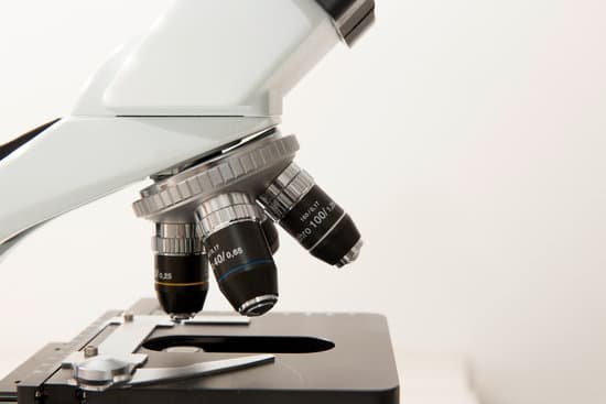
What does skeletal muscle look like under a microscope? Skeletal muscles are long and cylindrical in appearance; when viewed under a microscope, skeletal muscle tissue has a striped or striated appearance. The striations are caused by the regular arrangement of contractile proteins (actin and myosin). … Skeletal muscle also has multiple nuclei present in a single cell.
What do skeletal muscles look like when observed under a microscope? Skeletal muscle looks striped or “striated” – the fibres contain alternating light and dark bands (striations) like horizontal stripes on a rugby shirt. In skeletal muscle, the fibres are packed into regular parallel bundles.
How does a skeletal muscle look like? What do skeletal muscles look like? Skeletal muscle fibers are red and white. They look striated, or striped, so they’re often called striated muscles. Cardiac muscles are also striated, but smooth muscles aren’t.
What is the microscopic anatomy of skeletal muscle? The plasma membrane of the skeletal muscle fiber is called a sarcolemma. The muscle fiber contains long cylindrical structures, the myofibrils. The myofibrils almost entirely fill the cell and push the nuclei to the outer edges of the cell under the sarcolemma.
What does skeletal muscle look like under a microscope? – Related Questions
Is dic electron microscope?
D.I.C. and related optics give a specimen a three dimensional appearance that is not unlike the appearance of a specimen in a scanning electron microscope. These methods enhance depth of focus so that thicker specimens can be observed at higher magnifications.
How much can the most powerful light microscope magnify?
On a stock, high-performance compound light microscope, magnification levels of 1000x can be achieved (10x ocular lens, 100x objective lens). With that said, the maximum magnification level of a light microscope at the high end of the performance spectrum is 2000x magnification (20x ocular, 100x objective).
Which microscope lens provides the most magnification?
The oil immersion objective lens provides the most powerful magnification, with a whopping magnification total of 1000x when combined with a 10x eyepiece.
What is body tube in compound microscope?
The microscope body tube separates the objective and the eyepiece and assures continuous alignment of the optics. It is a standardized length, anthropometrically related to the distance between the height of a bench or tabletop (on which the microscope stands) and the position of the seated observer’s eyes.
How to not scratch lens on microscope?
Place the objective lens on a dust-free surface. 2. Gently blow away loose dust that is on the surface of the optical glass with a dust blower, as if any dust left on throughout the cleaning process could scratch the optical glass or coating. Blow the air across the lens surface to avoid damaging it.
How to perform a sperm count using a microscope?
Use the sterile dropper to place a drop of ejaculate onto a clean slide. Prepare the slide by placing a cover slip over the specimen. Count the sperm in the 400x field of view. Record the numbers on the analysis sheet, or multiply the number by .
What is na on a microscope?
Numerical aperture (abbreviated as ‘NA’) is an important consideration when trying to distinguish detail in a specimen viewed down the microscope. NA is a number without units and is related to the angles of light which are collected by a lens.
What is the best magnification for viewing blood stereo microscope?
– Is standard 400x magnification okay, or do you need 1000x magnification to see greater cell detail? 400x is ideal for high school biology; 1000x is best for college microbiology.
Which lens is used to construct a microscope?
The simplest compound microscope is constructed from two convex lenses as shown schematically in Figure 2. The first lens is called the objective lens, and has typical magnification values from 5× to 100×.
How do you regulate the diaphragm on a microscope?
Below. The light source is housed in the base of the microscope. It passes through the field iris diaphragm. The size of the field diaphragm is controlled by rotating a knurled ring which is concentric with it.
What is the function of mechanical stage in microscope?
All microscopes are designed to include a stage where the specimen (usually mounted onto a glass slide) is placed for observation. Stages are often equipped with a mechanical device that holds the specimen slide in place and can smoothly translate the slide back and forth as well as from side to side.
What is a microscope c mount?
C-Mount adapters are microscope specific, which means they are designed specifically for the brand of microscope to keep the camera in focus while the eyepieces are in focus. For example, if you are using a Zeiss microscope, you need to use the corresponding Zeiss c-mount adapter made for that microscope.
Where is the substage condenser on a microscope?
A microscope condenser, also called a substage condenser when it’s not above the stage (as in an inverted microscope), is defined as an optical lens which renders a divergent beam from a point source into a parallel or converging beam to illuminate an object.
How to figure out the magnification of a microscope?
To figure the total magnification of an image that you are viewing through the microscope is really quite simple. To get the total magnification take the power of the objective (4X, 10X, 40x) and multiply by the power of the eyepiece, usually 10X.
What are tem and sem microscopes?
The main difference between SEM and TEM is that SEM creates an image by detecting reflected or knocked-off electrons, while TEM uses transmitted electrons (electrons that are passing through the sample) to create an image.
Can to much motrin cause microscopic blood in urine?
Yes, ibuprofen can cause hematuria (blood in the urine). Due to you having blood in your urine it would most likely be recommended that you do not take ibuprofen or other NSAID in the future, unless you have been prescribed them. Other non-steroidal anti-inflammatory (NSAID) drugs may cause the same side effect.
What is compound microscope magnification at scanning power?
A scanning objective lens provides the lowest magnification power of all objective lenses. 4x is a common magnification for scanning objectives and, when combined with the magnification power of a 10x eyepiece lens, a 4x scanning objective lens gives a total magnification of 40x.
How to focus high power microscope camera?
Look at the objective lens (3) and the stage from the side and turn the focus knob (4) so the stage moves upward. Move it up as far as it will go without letting the objective touch the coverslip. Look through the eyepiece (1) and move the focus knob until the image comes into focus.
Can you see an ovum without a microscope?
Most cells aren’t visible to the naked eye: you need a microscope to see them. The human egg cell is an exception, it’s actually the biggest cell in the body and can be seen without a microscope. … That may sound small, but no other cell comes close to being that large.
Can you see dna under light microscope?
While it is possible to see the nucleus (containing DNA) using a light microscope, DNA strands/threads can only be viewed using microscopes that allow for higher resolution.
What microscope is used to see mycobacterium tuberculosis?
Because of the high incidence of death due to tuberculosis, Mycobacterium tuberculosis was one of the first bacterial species to be examined using the technique of electron microscopy (EM) [1],[2].

