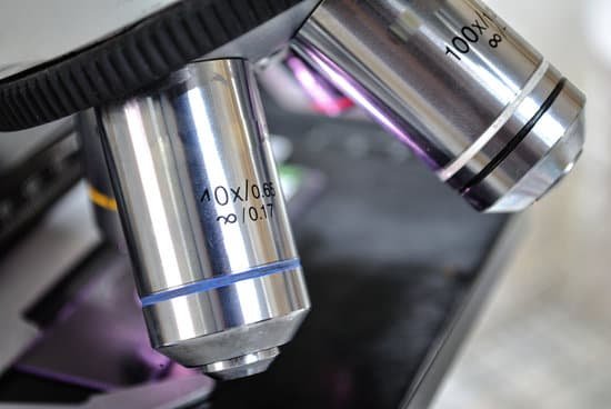
What does the ocular lens of a microscope do? The eyepiece, or ocular lens, is the part of the microscope that magnifies the image produced by the microscope’s objective so that it can be seen by the human eye.
What do the objective and ocular lenses do? The objective and ocular lenses are responsible for magnifying the image of the specimen being viewed. So for 10X objective and 10X ocular, … The value for numerical aperture measures to what extent the light that passes through a specimen is spread out over and collected by the objective lens.
What is meant by ocular lens? Lens, ocular: In a microscope, the lens closest to the eye. Also known as eyepiece. Most light microscopes are binocular, with one ocular lens for each eye.
What magnification do you need to see pinworms? 2. Observe the specimen, cover slip side up, under the microscope at 500X total magnification. Since egg production is variable, specimens should be taken on successive mornings. Three specimens should be sufficient for diagnosis.
What does the ocular lens of a microscope do? – Related Questions
What does a diaphragm on a microscope mean?
Iris Diaphragm controls the amount of light reaching the specimen. It is located above the condenser and below the stage. Most high quality microscopes include an Abbe condenser with an iris diaphragm. Combined, they control both the focus and quantity of light applied to the specimen.
How to estimate cell size under a microscope?
Divide the number of cells in view with the diameter of the field of view to figure the estimated length of the cell. If the number of cells is 50 and the diameter you are observing is 5 millimeters in length, then one cell is 0.1 millimeter long. Measured in microns, the cell would be 1,000 microns in length.
How small can an electron microscope see?
Light microscopes let us look at objects as long as a millimetre (10-3 m) and as small as 0.2 micrometres (0.2 thousands of a millimetre or 2 x 10⁻⁷ m), whereas the most powerful electron microscopes allow us to see objects as small as an atom (about one ten-millionth of a millimetre or 1 angstrom or 10-10 m).
Why are mechanical parts of the microscope important?
It is important to use the mechanical stage to get a better and clear view of the specimen. It makes using the microscope much easier. It allows for better control of the slide in addition to avoiding accidental bumping that may knock the slide out of focus.
What microscopic bugs crawl on skin?
The human itch mite (Sarcoptes scabiei var. hominis) is a microscopic bug that is one of the few to actually burrow and live beneath human skin. Adult female itch mites burrow under the top layer of skin, where they can continue to live and lay eggs for weeks undetected.
How do fibers look different under a microscope?
Under the microscope, the wool fiber looks like a long cylinder with scales on it. The fiber is very curly and springy. Cloth made from wool includes cashmere, camel’s hair, alpaca, covert cloth, flannel, gabardine, mohair, serge, tweed and worsted. … Cotton fibers are the hairs found on the seeds of the cotton plant.
Are dust mites microscopic?
Dust mites are microscopic, insect-like pests that generate some of the most common indoor substances—or allergens—that can trigger allergic reactions and asthma in many people. Hundreds of thousands of dust mites can live in the bedding, mattresses, upholstered furniture, carpets or curtains in your home.
What is microscope slide science?
A microscope slide is a thin flat piece of glass, typically 75 by 26 mm (3 by 1 inches) and about 1 mm thick, used to hold objects for examination under a microscope. Typically the object is mounted (secured) on the slide, and then both are inserted together in the microscope for viewing.
What was the first type of microscope developed?
A Dutch father-son team named Hans and Zacharias Janssen invented the first so-called compound microscope in the late 16th century when they discovered that, if they put a lens at the top and bottom of a tube and looked through it, objects on the other end became magnified.
How should a microscope be carried?
Always keep your microscope covered when not in use. Always carry a microscope with both hands. Grasp the arm with one hand and place the other hand under the base for support.
How much is a microscope worth?
The most popular compound microscopes from some of the most well-known brands cost on average around $900-$1,200, although there are beginner microscopes that are just above the toy level that cost $100.
Who invented the pocket microscope?
Benjamin Martin designed and built this microscope in the middle of the eighteenth century and named it the pocket microscope because of its small size and portability.
What is microscope and its parts?
The three basic, structural components of a compound microscope are the head, base and arm. Head/Body houses the optical parts in the upper part of the microscope. Base of the microscope supports the microscope and houses the illuminator. Arm connects to the base and supports the microscope head.
Which microscopes to use a beam of light?
Light microscopes use visible light which passes and bends through the lens system. Electron microscopes use a beam of electrons, opposed to visible light, for magnification.
Who invented the first digital microscope?
An early digital microscope was made by a lens company in Tokyo, Japan in 1986, which is now known as Hirox Co Ltd. It included a control box and a lens connected to a computer. Other versions of digital microscope were later developed by Keyence Corp and Leica Microsystems.
What is the difference between a microscope and a telescope?
Since telescopes view large objects — faraway objects, planets or other astronomical bodies — its objective lens produces a smaller version of the actual image. On the other hand, microscopes view very small objects, and its objective lens produces a larger version of the actual image.
How to look at semen under microscope?
You can view sperm at 400x magnification. You do NOT want a microscope that advertises anything above 1000x, it is just empty magnification and is unnecessary. In order to examine semen with the microscope you will need depression slides, cover slips, and a biological microscope.
How to get the magnification of a stereoscopic microscope?
On a basic stereo microscope setup, to determine total magnification simply look at the magnification on the eyepiece and on the zoom knob. Stereo microscope auxiliary lenses are only usually used when the working distance needs to be adjusted or in some cases if magnification is being pushed quite high.
What can i use to clean your microscope?
Put a small amount of lens cleaning fluid or cleaning mixture on the tip of the lens paper. We recommend 70% ethanol because it can effectively and safely clean and disinfect the surface. Larger surfaces, such as a glass plate, may be too large to wipe using this technique.
What is urinalysis microscopic on positives?
A routine Urinalysis with Microscopic Examination on Positives is used to detect abnormalities of urine; diagnose and manage renal diseases, urinary tract infection, urinary tract neoplasms, systemic diseases, and inflammatory or neoplastic diseases adjacent to the urinary tract.

