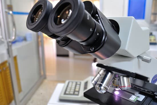
What does the stage opening do microscope? Stage opening – part of the mechanical stage that allows light to pass through the specimen for a better view. Stage lock control – the locking control that allows the user to fix the stage into position with respect to its rotation around the condenser axis.
What is the purpose of the hole in the stage? Stage Clips are used when there is no mechanical stage. The viewer is required to move the slide manually to view different sections of the specimen. Aperture is the hole in the stage through which the base (transmitted) light reaches the stage.
Does the stage move on a microscope? Your microscope slide should be prepared by placing a coverslip or cover glass over the specimen. This will help protect the objective lenses if they touch the slide. Place the microscope slide on the stage and fasten it with the stage clips. You can push down on the back end of the stage clip to open it.
Which microscope is better for magnification? Out of all types of microscopes, the electron microscope has the greatest capability in achieving high magnification and resolution levels, enabling us to look at things right down to each individual atom.
What does the stage opening do microscope? – Related Questions
Which is better in a microscope bigger or less resolution?
Using a larger monitor certainly can magnify the image further. But, it will still be just as blurry or sharp as the resolution. Fortunately, in general higher magnification lenses also have better resolution.
Why does the electron microscope have a higher resolving power?
Electron microscopes differ from light microscopes in that they produce an image of a specimen by using a beam of electrons rather than a beam of light. Electrons have much a shorter wavelength than visible light, and this allows electron microscopes to produce higher-resolution images than standard light microscopes.
What can you see with an electron microscope?
An electron microscope is a microscope that uses a beam of accelerated electrons as a source of illumination. … Electron microscopes are used to investigate the ultrastructure of a wide range of biological and inorganic specimens including microorganisms, cells, large molecules, biopsy samples, metals, and crystals.
How do modern light microscopes work?
A simple light microscope manipulates how light enters the eye using a convex lens, where both sides of the lens are curved outwards. When light reflects off of an object being viewed under the microscope and passes through the lens, it bends towards the eye. This makes the object look bigger than it actually is.
Is it possible to see atoms with a microscope?
Atoms are so small that it’s almost impossible to see them without microscopes. … The diameter of a strontium atom is a few millionths of a millimeter.
Is a microscope include with tympanoplasty?
Tympanoplasty was conventionally performed using a microscope for decades. However, since the endoscope began to be used in middle ear surgery in the 1970s, endoscopic tympanoplasty has gained increasing attention.
How to observe algae under microscope?
Place a drop of water or specimen on a microscope slide containing algae, gently cover the slide with a coverslip and view under a microscope. Be sure to use a specimen of very small quantity to avoid clumping. Observe slide in the microscopic range of about 40X or 100X magnification.
What is the function of the oculars on a microscope?
The eyepiece, or ocular lens, is the part of the microscope that magnifies the image produced by the microscope’s objective so that it can be seen by the human eye.
Why move the microscope condenser down?
If the diffuse surface of the illumination optics appears as a rough background when the specimen is in focus, move the condenser up and down so that the rough background is not visible.
What is the magnification of a transmission electron microscope?
Transmission electron microscopes (TEM) are microscopes that use a particle beam of electrons to visualize specimens and generate a highly-magnified image. TEMs can magnify objects up to 2 million times.
Do pathologists use electron microscopes?
The Diagnostic Electron Microscopy Unit provides diagnostic transmission electron microscopic services to the Mass General community, and to pathologists and researchers throughout the US and abroad.
What is the greatest contribution of microscope in biology?
The microscope is important because biology mainly deals with the study of cells (and their contents), genes, and all organisms. Some organisms are so small that they can only be seen by using magnifications of ×2000−×25000 , which can only be achieved by a microscope. Cells are too small to be seen with the naked eye.
Does temporal bone dissection use a microscope?
The temporal bone dissection procedure involves harvesting the bone from a human cadaver and storage in formaldehyde. It is mounted securely in a bone holder in the same position as in actual surgery and dissected using a surgical microscope and a micromotor drill.
Can you use a light compound microscope to see mitosis?
This is accomplished in a Eukaryote through mitosis. In this lab you will use the light microscope to view cells at different stages of mitosis as well as the division of the cell called cytokinesis. … Focusing the microscope with 40x objective should give you a close enough view of the chromosomes to find each phase.
What type of microscope is used to view ebola virus?
These steps in the replication cycle can be studied using electron microscopy (EM), including transmission electron microscopy (TEM) and scanning electron microscopy (SEM), which is one of the most useful methods for visualizing EBOV particles and EBOV-infected cells at the ultrastructural level.
What does a dissecting microscope?
A dissecting microscope is used to view three-dimensional objects and larger specimens, with a maximum magnification of 100x. This type of microscope might be used to study external features on an object or to examine structures not easily mounted onto flat slides.
How can fluorescence be used in microscopes?
Fluorescence microscopy is a technique whereby fluorescent substances are examined in a microscope. … The specimen is examined through a barrier filter that absorbs the short-wavelength light used for illumination and transmits the fluorescence, which is therefore seen as bright against a dark background (Figure 1).
What is the highest magnification on a light microscope?
Using the mathematical equations given above and the values for maximum numerical aperture attainable with the lenses of a light microscope it can be shown that the maximum useful magnification on a light microscope is between 1000X and 1500X. Higher magnification is possible, but resolution will not improve.
How worried should i be microscopic hematuria?
If the doctor rules out any medical problem causing hematuria, you will not need treatment. “If you find blood in your urine, or your doctor tells you that you have microscopic hematuria, don’t panic,” Dr.
What is the difference between macroscopic microscopic and submicroscopic?
As adjectives the difference between submicroscopic and macroscopic. is that submicroscopic is smaller than microscopic; too small to be seen even with a microscope while macroscopic is visible to the unassisted eye.

