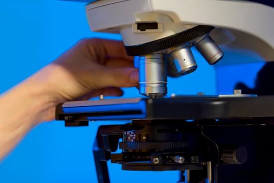
What does zoom magnification mean for microscope? Magnification is the ability of a microscope to produce an image of an object at a scale larger (or even smaller) than its actual size. … At the present time, magnification is well defined when viewing an image of a sample through the eyepieces of a microscope.
What is a good zoom for a microscope? Lower magnification (10-20x) produces a larger field of view and is best for young kids. It is also ideal for viewing stamps and coins. Higher magnification (30-40x) is better for close-ups and more detailed work.
What can you see with 1000x zoom microscope? At 1000x magnification you will be able to see 0.180mm, or 180 microns.
What is magnification zoom? Per Petteri, the term zoom refers to the ratio of the shortest focal length to its longest focal length, of a “zoom” (variable magnification) lens. For example, a 30mm – 300mm zoom lens will have a zoom ratio of 10 (300/30).
What does zoom magnification mean for microscope? – Related Questions
When not in use the microscope should be stored?
When the microscope is not in use keep it covered with the dust cover. This alone will extend the life of your microscope. Even if the microscope is stored within a cabinet, you should still cover it with the dust cover. Do not store a microscope without any eyepieces, even if it is covered.
What type of lens does a light microscope use?
Principles. The light microscope is an instrument for visualizing fine detail of an object. It does this by creating a magnified image through the use of a series of glass lenses, which first focus a beam of light onto or through an object, and convex objective lenses to enlarge the image formed.
Can you see sperm with a home microscope?
A semen microscope or sperm microscope is used to identify and count sperm. You can view sperm at 400x magnification. … You do NOT want a microscope that advertises anything above 1000x, it is just empty magnification and is unnecessary.
Do people all have microscopic bugs on their skin?
They’re known as Demodex or eyelash mites, and just about every adult human alive has a population living on them. The mostly transparent critters are too small to see with the naked eye.
What size would be considered microscopic?
So, we can think of the microscopic scale as being from a millimetre (10-3 m) to a ten-millionth of a millimetre (10-10 m). Even within the microscopic scale, there are immense variations in the size of objects.
How to measure field of view microscope?
For instance, if your eyepiece reads 10X/22, and the magnification of your objective lens is 40. First, multiply 10 and 40 to get 400. Then divide 22 by 400 to get a FOV diameter of 0.055 millimeters.
Who created the single lens microscope?
Dutch scientist Antoine van Leeuwenhoek designed high-powered single lens microscopes in the 1670s. With these he was the first to describe sperm (or spermatozoa) from dogs and humans.
What do live bacteria look like under a microscope?
In order to see bacteria, you will need to view them under the magnification of a microscopes as bacteria are too small to be observed by the naked eye. Most bacteria are 0.2 um in diameter and 2-8 um in length with a number of shapes, ranging from spheres to rods and spirals.
How do you determine the magnification power of a microscope?
To figure the total magnification of an image that you are viewing through the microscope is really quite simple. To get the total magnification take the power of the objective (4X, 10X, 40x) and multiply by the power of the eyepiece, usually 10X.
What are the three objective lenses of a compound microscope?
Most compound microscopes come with interchangeable lenses known as objective lenses. Objective lenses come in various magnification powers, with the most common being 4x, 10x, 40x, and 100x, also known as scanning, low power, high power, and (typically) oil immersion objectives, respectively.
How does a scanning probe microscope work?
How does it work? A scanning probe microscope has a sharp probe tip on the end of a cantilever, which can scan the surface of the specimen. The tip moves back and forth in a very controlled manner and it is possible to move the probe atom by atom.
What is an aperture on a microscope?
Numerical Aperture and Resolution. The numerical aperture of a microscope objective is the measure of its ability to gather light and to resolve fine specimen detail while working at a fixed object (or specimen) distance. … The smaller the object, the more pronounced the diffraction of incident light rays will be.
Why would scientist use electron microscope?
The electron microscope is an integral part of many laboratories. Researchers use it to examine biological materials (such as microorganisms and cells), a variety of large molecules, medical biopsy samples, metals and crystalline structures, and the characteristics of various surfaces.
What can be seen with the light microscope?
Explanation: You can see most bacteria and some organelles like mitochondria plus the human egg. You can not see the very smallest bacteria, viruses, macromolecules, ribosomes, proteins, and of course atoms.
What is the meaning of fine adjustment knob in microscope?
FINE ADJUSTMENT KNOB — A slow but precise control used to fine focus the image when viewing at the higher magnifications.
What is the microscopic agglutination test used for?
The microscopic agglutination test (MAT) is the gold standard for sero-diagnosis of leptospirosis because of its unsurpassed diagnostic specificity. It uses panels of live leptospires, ideally recent isolates, representing the circulating serovars from the area where the patient became infected.
What two parts do you hold when carrying a microscope?
When carrying a compound microscope always take care to lift it by both the arm and base, simultaneously. There are two optical systems in a compound microscope: Eyepiece Lenses and Objective Lenses: Eyepiece or Ocular is what you look through at the top of the microscope.
What does a condenser on a microscope do?
On upright microscopes, the condenser is located beneath the stage and serves to gather wavefronts from the microscope light source and concentrate them into a cone of light that illuminates the specimen with uniform intensity over the entire viewfield.
What does each part of a microscope do?
Eyepiece Lens: the lens at the top that you look through, usually 10x or 15x power. Tube: Connects the eyepiece to the objective lenses. Arm: Supports the tube and connects it to the base. Base: The bottom of the microscope, used for support.
How scanning transmission electron microscope works?
In the scanning transmission electron microscopy (STEM) mode, the microscope lenses are adjusted to create a focused convergent electron beam or probe at the sample surface. This focused probe is then scanned across the sample and various signals are collected point-by-point to form an image.
What do fluorescence microscopes look at?
A fluorescence microscope is an optical microscope that uses fluorescence instead of, or in addition to, scattering, reflection, and attenuation or absorption, to study the properties of organic or inorganic substances.

