What foods should you avoid if you have microscopic colitis? Avoid beverages that are high in sugar or sorbitol or contain alcohol or caffeine, such as coffee, tea and colas, which may aggravate your symptoms. Choose soft, easy-to-digest foods. These include applesauce, bananas, melons and rice. Avoid high-fiber foods such as beans and nuts, and eat only well-cooked vegetables.
Are bananas good for microscopic colitis? Bananas are a low-fiber fruit and many people who have colitis can easily digest them. When people who have colitis have flares (periods of worsening symptoms), they may find it helpful to eat bland foods, including bananas.
What foods trigger colitis? The outlook for people with Microscopic Colitis is generally good. Four out of five can expect to be fully recovered within three years, with some even recovering without treatment. However, for those who experience persistent or recurrent diarrhea, long term budesonide may be necessary.
How long does it take to recover from microscopic colitis? Some foods may bring on the condition in some people. Certain foods may also make lymphocytic colitis symptoms worse. These can include caffeine and milk products.
What foods should you avoid if you have microscopic colitis? – Related Questions
How many mm is the a microscope specimen?
A standard microscope slide measures about 75 mm by 25 mm (3″ by 1″) and is about 1 mm thick.
How many magnifying lenses are on a compound microscope?
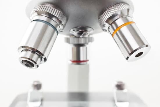
Generally there are 3 to 4 lenses in a compound microscope. Moreover, all these lenses have different power (magnification).
What is the definition of coarse adjustment on a microscope?
COARSE ADJUSTMENT KNOB — A rapid control which allows for quick focusing by moving the objective lens or stage up and down. It is used for initial focusing.
What was the first microscope used for?
1675: Enter Anton van Leeuwenhoek, who used a microscope with one lens to observe insects and other specimen. Leeuwenhoek was the first to observe bacteria.
Why is a vacuum needed in an electron microscope?
Most electron microscopes are high-vacuum instruments. Vacuums are needed to prevent electrical discharge in the gun assembly (arcing), and to allow the electrons to travel within the instrument unimpeded. … Also, any contaminants in the vacuum can be deposited upon the surface of the specimen as carbon.
What can be seen through an electron microscope?
An electron microscope is a microscope that uses a beam of accelerated electrons as a source of illumination. … Electron microscopes are used to investigate the ultrastructure of a wide range of biological and inorganic specimens including microorganisms, cells, large molecules, biopsy samples, metals, and crystals.
When is a light microscope used?
light microscopes are used to study living cells and for regular use when relatively low magnification and resolution is enough. electron microscopes provide higher magnifications and higher resolution images but cannot be used to view living cells.
Why is a light microscope called compound microscope?
The compound light microscope is a tool containing two lenses, which magnify, and a variety of knobs used to move and focus the specimen. Since it uses more than one lens, it is sometimes called the compound microscope in addition to being referred to as being a light microscope.
How to use scanning tunneling microscope?
The scanning tunneling microscope (STM) works by scanning a very sharp metal wire tip over a surface. By bringing the tip very close to the surface, and by applying an electrical voltage to the tip or sample, we can image the surface at an extremely small scale – down to resolving individual atoms.
How to prepare specimen for light microscope?
Preparation often involves nothing more than mounting a small piece of the specimen in a suitable liquid on a glass slide and covering it with a glass coverslip. ADVERTISEMENTS: The slide is then positioned on the specimen stage of the microscope and examined through the ocular lens, or with a camera.
What does the iris adjustment do on a microscope?
In light microscopy the iris diaphragm controls the size of the opening between the specimen and condenser, through which light passes. Closing the iris diaphragm will reduce the amount of illumination of the specimen but increases the amount of contrast.
When was the atom microscope invented?
The invention of the electron microscope by Max Knoll and Ernst Ruska at the Berlin Technische Hochschule in 1931 finally overcame the barrier to higher resolution that had been imposed by the limitations of visible light. Since then resolution has defined the progress of the technology.
Why do cork float in a microscope?
Wood and cork float for the same reason that life jackets and Styrofoam float: these materials have a lot of air in them, which makes them extremely buoyant. If you’ve ever looked at cork under the microscope, you’ve probably noticed it has a lot of holes. These holes trap air. It’s the same thing with wood and foam.
What type of microscopes are used to analyze fiber evidence?
Hair and fiber samples are often collected as trace evidence. Fiber types can be determined using optical microscopy techniques. Human hair can be distinguished from animal hair using a Scanning Electron Microscope (SEM).
Which microscope has the higher magnifying power?
Out of all types of microscopes, the electron microscope has the greatest capability in achieving high magnification and resolution levels, enabling us to look at things right down to each individual atom.
How does a microscope help scientists observe objects?
A microscope is an instrument that can be used to observe small objects, even cells. The image of an object is magnified through at least one lens in the microscope. This lens bends light toward the eye and makes an object appear larger than it actually is.
How small can a light microscope see?
The smallest thing that we can see with a ‘light’ microscope is about 500 nanometers. A nanometer is one-billionth (that’s 1,000,000,000th) of a meter. So the smallest thing that you can see with a light microscope is about 200 times smaller than the width of a hair.
Which part of the microscope controls the amount of light?
Iris diaphragm dial: Dial attached to the condenser that regulates the amount of light passing through the condenser. The iris diaphragm permits the best possible contrast when viewing the specimen.
Can you use a microscope to see sperm?
A semen microscope or sperm microscope is used to identify and count sperm. These microscopes are used when breeding animals or for examining human fertility. You can view sperm at 400x magnification.
How you handle a microscope properly?
Hold the microscope with one hand around the arm of the device, and the other hand under the base. This is the most secure way to hold and walk with the microscope. Avoid touching the lenses of the microscope. The oil and dirt on your fingers can scratch the glass.
How to choose a microscope objective?
What is the size of your specimen? Olympus’ microscope objectives feature a range of magnifications from 1.25x to 150x. This is the first parameter to consider when finding the best objective for your application. Combined with the magnification from the eyepieces, it determines the microscope’s overall magnification.

