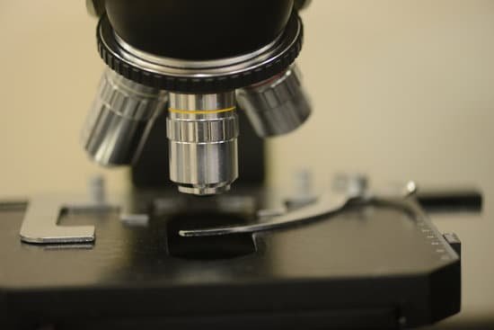
What is a diffuser on a microscope? A light diffuser for use in illumination systems of optical microscopes which utilize a light source to illuminate a specimen staged on the microscope for observation. … The light diffusing plate randomly breaks up light transmitted from a light source which passes therethrough into spatially isotopic light.
What is the function of diffuser in light? In optics, a diffuser (also called a light diffuser or optical diffuser) is any material that diffuses or scatters light in some manner to transmit soft light.
What is a holographic diffuser? Holographic diffusers (or diffractive homogenizers) are employed to control light distribution in illumination and laser systems. … Holographic diffusers are cost-effectively replicated in high volume using injection or compression molding or can be embossed onto surfaces of other optical components.
What is a light diffuser for photography? What Is a Light Diffuser? A light diffuser is a mechanism for scattering your light output. Light diffusion reduces harsh shadows and balances your lighting effects, creating even, soft light (like a lampshade) on your subjects.
What is a diffuser on a microscope? – Related Questions
What can be seen with an electron microscope?
An electron microscope is a microscope that uses a beam of accelerated electrons as a source of illumination. … Electron microscopes are used to investigate the ultrastructure of a wide range of biological and inorganic specimens including microorganisms, cells, large molecules, biopsy samples, metals, and crystals.
What has the microscope discovered?
Robert Hooke discovered cells by studying the honeycomb structure of a cork under a microscope. Marcello Marpighi, known as the father of microscopic anatomy, found taste buds and red blood cells. Robert Koch used a compound microscope to discover tubercle and cholera bacilli.
Are there microscopic black holes?
Micro black holes, also called quantum mechanical black holes or mini black holes, are hypothetical tiny black holes, for which quantum mechanical effects play an important role. … However, such quantum black holes would instantly evaporate, either totally or leaving only a very weakly interacting residue.
Why can’t i see through my microscope?
if you place a large and dark specimen on the stage, then the light of the microscope is not able to pass though the object. You will not be able to see anything except a dark shadow without much detail. In this case you must either cut the specimen into thin sections, tear it apart or squash it.
What is the function of oil immersion in microscope?
Immersion oil increases the resolving power of the microscope by replacing the air gap between the immersion objective lens and cover glass with a high refractive index medium and reducing light refraction.
When using microscopes the resolution refers to?
In microscopy, the term ‘resolution’ is used to describe the ability of a microscope to distinguish detail. In other words, this is the minimum distance at which two distinct points of a specimen can still be seen – either by the observer or the microscope camera – as separate entities.
How do you view mold under microscope?
You can identify this type of mold by making a slide and viewing it under the microscope. It has a thin branch-like structure, with heads that look like blooming flowers, and release spherical spores.
What does cytoplasm look like under a microscope?
The cytoplasm is granulated with tiny dots all over. Under a high power microscope, the cell organelles are more differentiated and allow the observation of individual structures. Because of the affinity of the stain with the DNA and RNA of the cell, the components inside the nucleus might also be visible.
Do fungi have cell walls microscope?
Two different structures of the fungal cell wall define two different categories of fungi. … Under a microscope, these hyphae appear as single long cells with many nuclei. Septate fungi form cell walls between the cells of their hyphae, called septa.
How to clean your microscope?
To clean the eyepiece lens of your microscope, breathe onto the eyepiece lens and then wipe with lens tissue. For dirt that is difficult to remove, add ethanol (methanol in extreme cases) to a cotton swab, wipe the surface and then dry with a dry swab.
How to get rid of microscopic ants?
If you’ve seen ants marching in a line, try wiping down the surface with vinegar or bleach to disrupt the chemical trail. Prevent ants from entering your home in the first place by sealing up cracks and holes in walls. This will also prevent them from nesting inside wall cavities.
What is phase contrast microscope in biology?
Unstained living cells absorb practically no light. Phase-contrast microscopy is an optical microscopy technique that converts phase shifts in the light passing through a transparent specimen to brightness changes in the image. …
Can pepto bismol treat microscopic colitis?
Microscopic colitis can get better on its own, but most patients have recurrent symptoms. The main treatment for microscopic colitis is medication. In many cases, the doctor will start treatment with an antidiarrheal medication such as Pepto-Bismol® or Imodium® .
When and who invented the compound microscope and electron microscope?
In the late 16th century several Dutch lens makers designed devices that magnified objects, but in 1609 Galileo Galilei perfected the first device known as a microscope. Dutch spectacle makers Zaccharias Janssen and Hans Lipperhey are noted as the first men to develop the concept of the compound microscope.
What is the lowest magnification on a microscope?
A scanning objective lens provides the lowest magnification power of all objective lenses. 4x is a common magnification for scanning objectives and, when combined with the magnification power of a 10x eyepiece lens, a 4x scanning objective lens gives a total magnification of 40x.
Why do we stain specimens under a microscope?
The most basic reason that cells are stained is to enhance visualization of the cell or certain cellular components under a microscope. Cells may also be stained to highlight metabolic processes or to differentiate between live and dead cells in a sample.
How to determine wavelength in electron microscope?
wavelength of an electron is calculated for a given energy (accelerating voltage) by using the de Broglie relation between the momentum p and the wavelength λ of an electron (λ=h/p, h is Planck constant).
Is microscopic blood in urine dangerous?
While in many instances the cause is harmless, blood in urine (hematuria) can indicate a serious disorder. Blood that you can see is called gross hematuria. Urinary blood that’s visible only under a microscope (microscopic hematuria) is found when your doctor tests your urine.
How are electron microscopes useful?
Electron microscopes are used to investigate the ultrastructure of a wide range of biological and inorganic specimens including microorganisms, cells, large molecules, biopsy samples, metals, and crystals. Industrially, electron microscopes are often used for quality control and failure analysis.
Can’t focus microscope objective?
The height of your condenser may be set too high or too low (this can also affect resolution). Make sure that your objective lenses are screwed all the way into the body of the microscope. On high school microscopes, if someone adjusts the rack stop, the microscope will not focus.
What are the uses of ultraviolet microscope?
Recent study shows how a microscope using ultraviolet (UV) light to illuminate samples enables pathologists to assess high-resolution images of biopsies and other fresh tissue samples for disease, quickly and effectively.

