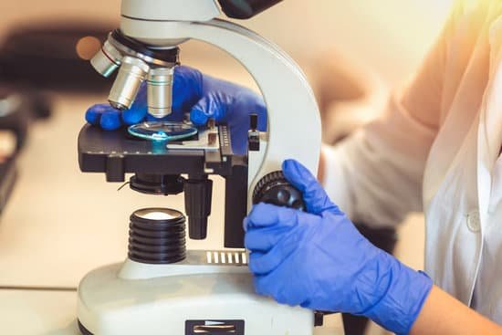
What is a microscope used for in a lab? The goal of any laboratory microscope is to produce clear, high-quality images, whether an optical microscope, which uses light to generate the image, a scanning or transmission electron microscope (using electrons), or a scanning probe microscope (using a probe).
What is a microscope and what is it used for? A microscope is an instrument that can be used to observe small objects, even cells. The image of an object is magnified through at least one lens in the microscope. This lens bends light toward the eye and makes an object appear larger than it actually is.
What kind of microscope is used in labs? The common light microscope used in the laboratory is called a compound microscope because it contains two types of lenses that function to magnify an object.
What are the three uses of microscope? They are used in different fields for different purposes. Some of their uses are tissue analysis, the examination of forensic evidence, to determine the health of the ecosystem, studying the role of protein within the cell, and the study of atomic structure.
What is a microscope used for in a lab? – Related Questions
How does a plant cell look like under the microscope?
Under the microscope, plant cells are seen as large rectangular interlocking blocks. The cell wall is distinctly visible around each cell. The cell wall is somewhat thick and is seen rightly when stained. The cytoplasm is also lightly stained containing a darkly stained nucleus at the periphery of the cell.
What does a prophase look like through a microscope?
The nuclear envelope breaks down, and the nucleolus disappears. The cytoskeleton also disassembles, and those microtubules form the spindle apparatus. When you look at a cell in prophase under the microscope, you will see thick strands of DNA loose in the cell.
What does a transmission electron microscope use?
Transmission electron microscopes (TEM) are microscopes that use a particle beam of electrons to visualize specimens and generate a highly-magnified image. TEMs can magnify objects up to 2 million times.
How did microscopes help scientists understand cells?
Microscopes allow humans to see cells that are too tiny to see with the naked eye. Therefore, once they were invented, a whole new microscopic world emerged for people to discover. … It allowed them to observe Eukaryotic cells with a nucleus and membrane-bound organelles that perform different life functions.
What does a diaphragm do microscope?
Opening and closing of the condenser aperture diaphragm controls the angle of the light cone reaching the specimen. The setting of the condenser’s aperture diaphragm, along with the aperture of the objective, determines the realized numerical aperture of the microscope system.
Which electron microscope produces a 3d image?
Scanning Electron Microscopes produce three-dimensional (3D) images while Transmission Electron Microscopes only produce flat (2D) images.
Who invented compound microscope?
Janssen was the son of a spectacle maker named Hans Janssen, in Middleburg, Holland, and while Zacharias is credited with inventing the compound microscope, most historians surmise that his father must have played a vital role, since Zacharias was still in his teens in the 1590s.
What is the similarities between light and electron microscopes?
Light microscopes and electron microscopes both use radiation – in the form of either light or electron beams, to form larger and more detailed images of objects (e.g. biological specimens, materials, crystal structures, etc.) than the human eye can produce unaided.
What is a microscope that can view atoms?
An electron microscope can be used to magnify things over 500,000 times, enough to see lots of details inside cells. There are several types of electron microscope. A transmission electron microscope can be used to see nanoparticles and atoms.
What is the lens of microscope?
A lens is an optical device that can reflect light. The reflection depends on the shape of a lens, which is typically convex or concave. For the purposes of microscopy, convex lenses are used for their ability to focus light at a single point.
What is effective focal length of microscope objective?
The focal length of a microscope objective is typically between 2 mm and 40 mm. However, that parameter is often considered as less important, since magnification and numerical aperture are sufficient for quantifying the essential performance in a microscope.
How has the microscope impacted medicine?
The microscope has had a major impact in the medical field. Doctors use microscopes to spot abnormal cells and to identify the different types of cells. This helps in identifying and treating diseases such as sickle cell caused by abnormal cells that have a sickle like shape.
What is the total lateral magnification of the microscope?
The total magnification of the microscope is calculated from the magnifying power of the objective multiplied by the magnification of the eyepiece and, where applicable, multiplied by intermediate magnifications.
Why are thing reversed and inverted under a light microscope?
The letter appears upside down and backwards because of two sets of mirrors in the microscope. This means that the slide must be moved in the opposite direction that you want the image to move. … These slides are thick, so they should only be viewed under low power.
Do microscopic organisms have eyes?
Scientists studying microscopic organisms in a seawater sample found a creature that appeared to be a tiny floating eyeball staring back at them. Although the creature is a single-celled organism, it possesses many of the features of the human eye, including a lens, cornea and retina.
Who made the modern microscope?
In the late 16th century several Dutch lens makers designed devices that magnified objects, but in 1609 Galileo Galilei perfected the first device known as a microscope. Dutch spectacle makers Zaccharias Janssen and Hans Lipperhey are noted as the first men to develop the concept of the compound microscope.
What do the objectives do on a microscope?
Objectives are responsible for primary image formation and play a central role in establishing the quality of images that the microscope is capable of producing. Furthermore, the magnification of a particular specimen and the resolution under which fine specimen detail also heavily depends on microscope objectives.
What microscope can see blood cells?
Compound microscopes magnify the tiny detail and structure of plant cells, bone marrow and blood cells, single-celled creatures like amoebas, and much more. Almost every homeschool family or hobbyist will need a 400x compound microscope to study cells and tiny organisms in biology and life science.
How are lenses used in telescopes microscopes and cameras?
They use mirrors and lenses to reflect and refract light and form images. The light microscope and telescope use convex lenses and mirrors to make enlarged images of very tiny or distant objects. A camera uses a convex lens to make a reduced image of an object.
Can viruses be seen with a brightfield microscope?
However, these microscopes are limited by light itself as they cannot show anything smaller than half the wavelength of visible light – and viruses are much smaller than this. But we can use microscopes to see the damage viruses do to our cells.
What is the difference between macroscopic microscopic and particulate?
The physical properties of matter can be viewed from either the macroscopic and microscopic level. The macroscopic level includes anything seen with the naked eye and the microscopic level includes atoms and molecules, things not seen with the naked eye. Both levels describe matter.

