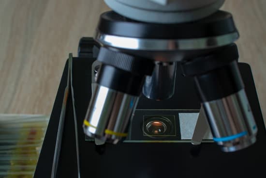
What is a microscope used to observe? A microscope is an instrument that can be used to observe small objects, even cells. The image of an object is magnified through at least one lens in the microscope. This lens bends light toward the eye and makes an object appear larger than it actually is.
What microscopes are used to observe cells? Two types of electron microscopy—transmission and scanning—are widely used to study cells. In principle, transmission electron microscopy is similar to the observation of stained cells with the bright-field light microscope.
What limits the resolving power of all microscopes quizlet? Wavelength is the main limiting factor on resolution because the image of two particles cannot be seen individually if it is smaller than the wavelength.
Why is there a limit to the resolving power of a compound microscope? In a compound microscope, the wavelength of the light waves that illuminate the specimen limits the resolution. The wavelength of visible light ranges from about 400 to 700 nanometers. The best compound microscopes cannot resolve parts of a specimen that are closer together than about 200 nanometers.
What is a microscope used to observe? – Related Questions
How to adjust eyepiece on a microscope?
This is very simple – most microscopes have an adjuster wheel in the centre of the eyepieces to adjust the distance. Otherwise, slide the eyepiece housing to match the width of your eyes. Once you have set this distance, you can then make the diopter adjustment.
What do all the parts of a microscope do?
All of the parts of a microscope work together – The light from the illuminator passes through the aperture, through the slide, and through the objective lens, where the image of the specimen is magnified.
Can you see cyclosis under the microscope?
In Elodea, cyclosis is easy to observe because chloroplasts move with the cytoplasm as it flows. … Tungsten or halogen substage microscope lamps produce both heat and light, so after 2–3 minutes, students should be able to observe the movement of chloroplasts.
What is the difference between microscopic and macroscopic?
The macroscopic level includes anything seen with the naked eye and the microscopic level includes atoms and molecules, things not seen with the naked eye. Both levels describe matter.
Who invented interference microscope?
The basic differential interference contrast (DIC) system, first devised by Francis Smith in 1955, is a modified polarized light microscope with two Wollaston prisms added, one to the front focal plane of the condenser and a second at the rear focal plane of the objective (see Figure 1).
Why use a stereoscopic microscope?
A stereo microscope is used for low-magnification applications, allowing high-quality, 3D observation of subjects that are normally visible to the naked eye. In life science stereo microscope applications, this could involve the observation of insects or plant life.
What is the mechanical stage of a microscope used for?
This mechanical stage contains controls for right-handed microscopists that allow the movement of the specimen slide in both the X (right and left) and Y (back and forth) directions so the microscopist can examine the entire microscope slide (secured to the stage with the slide holder).
Why is the specimen inverted in the microscope?
Under the slide on which the object is being magnified, there is a light source that shines up and helps you to see the object better. This light is then refracted, or bent around the lens. Once it comes out of the other side, the two rays converge to make an enlarged and inverted image.
What can you eat with microscopic colitis?
Avoid beverages that are high in sugar or sorbitol or contain alcohol or caffeine, such as coffee, tea and colas, which may aggravate your symptoms. Choose soft, easy-to-digest foods. These include applesauce, bananas, melons and rice. Avoid high-fiber foods such as beans and nuts, and eat only well-cooked vegetables.
What are these tiny microscopic red bugs?
Clover mites are very small, which is why they are often referred to as those tiny red bugs. The adults are reddish to brown in color and the immature mites and eggs are a bright red. Clover mites have eight legs with two at the head that are often thought to be antennae, not that you can see them that well.
How to label microscope slides?
We should say that the first thing to write upon a label is the genus and species of the plant; the next thing would be the name of the organ or tissue, and then might be added the date of collection, e.g., Marchanita polymorpha, young archegonia, January 10, 1915.
Why is the scanning tunneling microscope important?
Scanning Tunneling Microscopy allows researchers to map a conductive sample’s surface atom by atom with ultra-high resolution, without the use of electron beams or light, and has revealed insights into matter at the atomic level for nearly forty years.
What is the highest magnification on a compound microscope?
Typically, a compound microscope is used for viewing samples at high magnification (40 – 1000x), which is achieved by the combined effect of two sets of lenses: the ocular lens (in the eyepiece) and the objective lenses (close to the sample).
What are the four types of microscopes?
There are several different types of microscopes used in light microscopy, and the four most popular types are Compound, Stereo, Digital and the Pocket or handheld microscopes.
How does a simple light microscope work?
A simple light microscope manipulates how light enters the eye using a convex lens, where both sides of the lens are curved outwards. When light reflects off of an object being viewed under the microscope and passes through the lens, it bends towards the eye. This makes the object look bigger than it actually is.
What does the iris diaphragm on a microscope?
Iris Diaphragm controls the amount of light reaching the specimen. It is located above the condenser and below the stage. Most high quality microscopes include an Abbe condenser with an iris diaphragm. Combined, they control both the focus and quantity of light applied to the specimen.
What is the simple microscope made of?
A simple microscope is essentially a magnifying glass made of a single convex lens with a short focal length, which magnifies the object through angular magnification, thus producing an erect virtual image of the object near the lens.
Why use gold particles in electron microscope?
Gold is used for its high electron density which increases electron scatter to give high contrast ‘dark spots’. First used in 1971, immunogold labeling has been applied to both transmission electron microscopy and scanning electron microscopy, as well as brightfield microscopy.
Was invented microscope?
In the late 16th century several Dutch lens makers designed devices that magnified objects, but in 1609 Galileo Galilei perfected the first device known as a microscope.
Can genes be seen under a microscope?
Chromosomes, the spiraling strands of DNA that package the series of chemical bits called genes, are easily visible through a strong enough microscope if the right stain is used. … Chromosomes are best seen at the point in cell division called the metaphase stage of mitosis.

