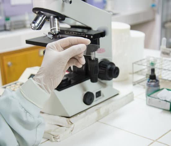
What is an electron microscope biology? Electron microscopy (EM) is a technique for obtaining high resolution images of biological and non-biological specimens. It is used in biomedical research to investigate the detailed structure of tissues, cells, organelles and macromolecular complexes.
What is an electron microscope GCSE? Electron microscopes use a beam of electrons instead of beams or rays of light. Living cells cannot be observed using an electron microscope because samples are placed in a vacuum.
What microscope is used in biology? Compound microscopes are designed with two or more convex lenses, allowing a high magnification level. This microscope allows the user to see detailed micro-organisms, bacteria, blood samples, cells and tissues. Ideally the compound microscope is used in scientific, medical research, education and biology applications.
What do electron microscopes see? Electron microscopy uses electrons to “see” small objects in the same way that light beams let us observe our surroundings or objects in a light microscope. With EM, we can look at the feather-like scales of an insect, the internal structures of a cell, individual proteins or even individual atoms in a metal alloy.
What is an electron microscope biology? – Related Questions
Can you see chlamydia under microscope?
The discharge is usually clear and stringy. In a sexual health clinic, the doctor or nurse may take a specimen and look at this under the microscope. They are looking for signs of infection such as an increased amount of white blood cells, and the chlamydia bacteria.
Which knobs move the slides on a microscope?
While observing through the eyepiece while focusing, move the mechanical stage, and thus the slide containing the specimen using the X knob, which moves the stage right and left, and the Y knob, which moves the stage back and forth.
What does the objective lens do on the microscope?
The objective, located closest to the object, relays a real image of the object to the eyepiece. This part of the microscope is needed to produce the base magnification. The eyepiece, located closest to the eye or sensor, projects and magnifies this real image and yields a virtual image of the object.
What are the parts of microscope function?
Eyepiece Lens: the lens at the top that you look through, usually 10x or 15x power. Tube: Connects the eyepiece to the objective lenses. Arm: Supports the tube and connects it to the base. Base: The bottom of the microscope, used for support. Illuminator: A steady light source (110 volts) used in place of a mirror.
Can an atom be seen with an electron microscope?
Using electron microscopes, it is possible to image individual atoms. Summary: Scientists have calculated how it is possible to look inside the atom to image individual electron orbitals. An electron microscope can’t just snap a photo like a mobile phone camera can.
What is diopter adjustment in microscope?
Diopter Adjustment: When you look through a microscope with two eyepiece lenses, you must be able to change the focus on one eyepiece to compensate for the difference in vision between your two eyes.
What microscopic bugs live on the human body?
Speaking of mites that feed on human material, Demodex folliculorum (Simon) is one of three mite species living on your face. The microscopic critters are found across the human body, but are particularly dense near the nose, eyebrows and eyelashes.
What is the use of microscope in laboratory apparatus?
A microscope is an instrument that is used to magnify small objects. Some microscopes can even be used to observe an object at the cellular level, allowing scientists to see the shape of a cell, its nucleus, mitochondria, and other organelles.
Which parts of the microscope magnify?
They have an objective lens (which sits close to the object) and an eyepiece lens (which sits closer to your eye). Both of these contribute to the magnification of the object.
What does the lamp do on a microscope?
lamp – produces the light (Typically, lamps are tungsten-filament light bulbs. For specialized applications, mercury or xenon lamps may be used to produce ultraviolet light. Some microscopes even use lasers to scan the specimen.)
How to determine field of view microscope?
For instance, if your eyepiece reads 10X/22, and the magnification of your objective lens is 40. First, multiply 10 and 40 to get 400. Then divide 22 by 400 to get a FOV diameter of 0.055 millimeters.
What causes microscopic hematuria in urine?
The most common causes of microscopic hematuria are urinary tract infection, benign prostatic hyperplasia, and urinary calculi. However, up to 5% of patients with asymptomatic microscopic hematuria are found to have a urinary tract malignancy.
How expensive electron microscope?
The price of a new electron microscope can range from $80,000 to $10,000,000 depending on certain configurations, customizations, components, and resolution, but the average cost of an electron microscope is $294,000. The price of electron microscopes can also vary by type of electron microscope.
Can you see dna with an electron microscope?
To view the DNA as well as a variety of other protein molecules, an electron microscope is used. … This is achieved because electron microscopes use electron beams rather than the visible light used for light microscopes.
Can aspirin cause microscopic blood in urine?
Certain medications including aspirin, heparin or warfarin (a blood thinner) and anti-cancer drug cyclophosphamide have been known to cause blood in urine. Sometimes, it’s just tiny traces that can only be seen under a microscope. This is called microscopic hematuria.
What are the three type of super resolution microscopes?
There are three main types of super-resolution microscopy, each one working via a different mechanism. These include stimulated emission depletion (STED) microscopy, structured illumination microscopy (SIM), and stochastic optical reconstruction microscopy (STORM)/photoactivation localization microscopy (PALM).
What does the eyepiece lens do on a microscope?
The eyepiece, or ocular lens, is the part of the microscope that magnifies the image produced by the microscope’s objective so that it can be seen by the human eye.
What is a scanning electron microscope used to observe?
Scanning electron microscope (SEM) is used to study the topography of materials and has a resolution of ∼2 nm. An electron probe is scanning over the surface of the material and these electrons interact with the material. Secondary electrons are emitted from the surface of the specimen and recorded.
What is a scanning electron microscope what are its advantages?
Advantages of a Scanning Electron Microscope include its wide-array of applications, the detailed three-dimensional and topographical imaging and the versatile information garnered from different detectors. … In addition, the technological advances in modern SEMs allow for the generation of data in digital form.
What is a fluorescence microscope use a fluorescent light?
A fluorescence microscope, on the other hand, uses a much higher intensity light source which excites a fluorescent species in a sample of interest. This fluorescent species in turn emits a lower energy light of a longer wavelength that produces the magnified image instead of the original light source.
What is a correct description of a confocal microscope quizlet?
Only $35.99/year. Confocal microscope. 1. visualizes fluorescent molecules in a single plane of focus by excluding out of focus light.

