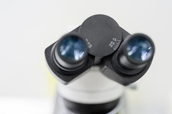
What is arm in microscope? Arm – The arm of the microscope supports the body tube. Body Tube – The body tube is a hollow tube through which light travels from the objective to the ocular. It contains a prism at the base of the tube that bends the light rays so they can enter the inclined tube.
What is the function of the arm in a light microscope? Arm: Supports the tube and connects it to the base. Base: The bottom of the microscope, used for support. Illuminator: A steady light source (110 volts) used in place of a mirror. If your microscope has a mirror, it is used to reflect light from an external light source up through the bottom of the stage.
What is a fixed arm on a microscope? Fixed Arm: A type of stand used with low power stereo microscopes. The arm and body are integral parts of the microscope and connected solidly to the base.
What is the function of the condenser in a bright field microscope? Most microscopes will have a built-in illuminator. The condenser is used to focus light on the specimen through an opening in the stage. After passing through the specimen, the light is displayed to the eye with an apparent field that is much larger than the area illuminated.
What is arm in microscope? – Related Questions
Why would you decrease amount of light with a microscope?
The light intensity decreases as magnification increases. There is a fixed amount of light per area, and when you increase the magnification of an area, you look at a smaller area. So you see less light, and the image appears dimmer. Image brightness is inversely proportional to the magnification squared.
What can a compound light microscope see?
With higher levels of magnification than stereo microscopes, a compound microscope uses a compound lens to view specimens which cannot be seen at lower magnification, such as cell structures, blood, or water organisms.
How is light polarized in a petrographic microscope?
There are usually two polarizing filters: the polarizer and the analyzer. The polarizer is located below the specimen stage and can be rotated through 360°. It helps to polarize the light which falls on the specimen. The analyzer is placed above the objective and may be rotatable in some cases.
Which type of microscope shows cells against a bright background?
Under the brightfield microscope, the technician can barely see the bacteria cells because they are nearly transparent against the bright background.
How to focus a microscope safely?
To focus a microscope, rotate to the lowest-power objective, and place your sample under the stage clips. Play with the magnification using the coarse adjustment knob and move your slide around until it is centered.
What is the purpose of the electron microscope?
Electron microscopy (EM) is a technique for obtaining high resolution images of biological and non-biological specimens. It is used in biomedical research to investigate the detailed structure of tissues, cells, organelles and macromolecular complexes.
What does a fine focus do on a microscope?
Focus (fine), Use the fine focus knob to sharpen the focus quality of the image after it has been brought into focus with the coarse focus knob. Illuminator, There is an illuminator built into the base of most microscopes.
Is it common to have microscopic blood in urine?
Microscopic hematuria, a common finding on routine urinalysis of adults, is clinically significant when three to five red blood cells per high-power field are visible. Etiologies of microscopic hematuria range from incidental causes to life-threatening urinary tract neoplasm.
Who invented microscope for the first time?
Lens Crafters Circa 1590: Invention of the Microscope. Every major field of science has benefited from the use of some form of microscope, an invention that dates back to the late 16th century and a modest Dutch eyeglass maker named Zacharias Janssen.
What is the significance of the compound microscope?
Compound microscopes can magnify specimens enough so that the user can see cells, bacteria, algae, and protozoa. You cannot see viruses, molecules, or atoms using a compound microscope because they are too small; an electron microscope is necessary to image such things.
Where was the electron microscopes invented?
The invention of the electron microscope by Max Knoll and Ernst Ruska at the Berlin Technische Hochschule in 1931 finally overcame the barrier to higher resolution that had been imposed by the limitations of visible light. Since then resolution has defined the progress of the technology.
Which microscope can magnify up to a million times?
What is a Transmission Electron Microscope? Transmission electron microscopes (TEM) are microscopes that use a particle beam of electrons to visualize specimens and generate a highly-magnified image. TEMs can magnify objects up to 2 million times.
Who first used the microscope to observe bacteria?
Antonie van Leeuwenhoek used single-lens microscopes, which he made, to make the first observations of bacteria and protozoa.
What type of microscope is used to view diseases?
Electron microscopy (EM) has long been used in the discovery and description of viruses. Organisms smaller than bacteria have been known to exist since the late 19th century (11), but the first EM visualization of a virus came only after the electron microscope was developed.
Who viewed cork under a microscope?
The first person to observe cells was Robert Hooke. Hooke was an English scientist. He used a compound microscope to look at thin slices of cork. Cork is found in some plants.
Why are light microscopes limited to 1000x?
The maximum magnification power of optical microscopes is typically limited to around 1000x because of the limited resolving power of visible light. … Modified environments such as the use of oil or ultraviolet light can increase the magnification.
What is ocular tube on microscope?
Eyepiece Tube holds the eyepieces in place above the objective lens. … Standard objectives include 4x, 10x, 40x and 100x although different power objectives are available. Coarse and Fine Focus knobs are used to focus the microscope.
What does the condenser do to the microscope?
On upright microscopes, the condenser is located beneath the stage and serves to gather wavefronts from the microscope light source and concentrate them into a cone of light that illuminates the specimen with uniform intensity over the entire viewfield.
What is the use of iris diaphragm in microscope?
Iris Diaphragm controls the amount of light reaching the specimen. It is located above the condenser and below the stage. Most high quality microscopes include an Abbe condenser with an iris diaphragm. Combined, they control both the focus and quantity of light applied to the specimen.
How much does it cost to use an electron microscope?
A typical commercial transmission electron microscope (TEM) costs about $2 for each electron volt of energy in the beam, and if you add on all the options, it can cost about $4–5 per eV. As you’ll see, we use beam energies in the range from 100,000–400,000 eV, so a TEM becomes an extremely expensive piece of equipment.

