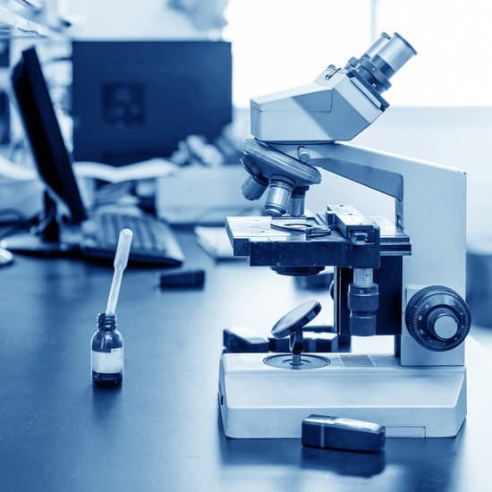
What is cmos microscope? Most microscope photodetector systems have some form of charged coupled device (CCD) or complementary metal oxide semiconductor (CMOS) as the image sensor. … During image capture, charge builds up in each photodiode (pixel). Image capture is then ended, and the information is read out sequentially.
What is a CMOS sensor used for? A CMOS sensor is an electronic chip that converts photons to electrons for digital processing. CMOS (complementary metal oxide semiconductor) sensors are used to create images in digital cameras, digital video cameras and digital CCTV cameras.
How does a CMOS sensor work? In a CMOS sensor, the charge from the photosensitive pixel is converted to a voltage at the pixel site and the signal is multiplexed by row and column to multiple on chip digital-to-analog converters (DACs). Inherent to its design, CMOS is a digital device.
What is difference between CMOS and CCD? One difference between CCD and CMOS sensors is the way they capture each frame. A CCD uses what’s called a “Global Shutter” while CMOS sensors use a “Rolling Shutter”. Global Shutter means that the entire frame is captured at the exact same time. … A CMOS sensor captures light though capturing each pixel one-by-one.
What is cmos microscope? – Related Questions
Does a microscope see in 2d or 3d?
Stereo 3D microscopes produce real-time 3D images, but they are usually limited to low-magnification applications, such as dissection. Most compound light microscopes produce flat, 2D images because high-magnification microscope lenses have inherently shallow depth of field, rendering most of the image out of focus.
What does bacteria look like under microscope?
In order to see bacteria, you will need to view them under the magnification of a microscopes as bacteria are too small to be observed by the naked eye. Most bacteria are 0.2 um in diameter and 2-8 um in length with a number of shapes, ranging from spheres to rods and spirals.
What to do when using a microscope?
Look through the eyepiece (1) and move the focus knob until the image comes into focus. Adjust the condenser (7) and light intensity for the greatest amount of light. Move the microscope slide around until the sample is in the centre of the field of view (what you see).
Can i use multispectral imaging equipment on a microscope?
The multispectral camera can be coupled to a compound microscope (left) and used to observe slide mounts of pigments (top centre). Some grains of pigments can be extracted from the surface of a painting or even from a layer of paint in a cross-section sample and can be mounted with a resin on a microscope slide.
Can atoms be seen with powerful microscopes?
Atoms are really small. So small, in fact, that it’s impossible to see one with the naked eye, even with the most powerful of microscopes. … Now, a photograph shows a single atom floating in an electric field, and it’s large enough to see without any kind of microscope.
What is the difference between light microscope and electron microscope?
Electron microscopes differ from light microscopes in that they produce an image of a specimen by using a beam of electrons rather than a beam of light. Electrons have much a shorter wavelength than visible light, and this allows electron microscopes to produce higher-resolution images than standard light microscopes.
What does salt look like under a 40x microscope?
Cube shaped salt crystals under 40x magnification. … The salt crystals are clearly cubic, even though some of the grains seem to be made up of overlapping cubes. The atoms that make up salt’s atomic lattice are arranged in a cubic shape, which results in the shape of the salt crystals.
What is a single lens microscope called?
The simple microscope consists of a single lens traditionally called a loupe. The most familiar present-day example is a reading or magnifying glass. Present-day higher-magnification lenses are often made with two glass elements that produce a colour-corrected image.
Who invented the microscope during the scientific revolution?
During the scientific revolution, Janssen invented a microscope and this instrument helped others study the natural world. This also lead to new discoveries. Janssen’s invention was a huge advancement in technology at that time.
Is telescope and microscope magnification the same?
In both the telescope and the microscope, the eyepiece magnifies the intermediate image; in the telescope, however, this is the only magnification.
What is numerical aperture of a microscope?
Numerical Aperture and Resolution. The numerical aperture of a microscope objective is the measure of its ability to gather light and to resolve fine specimen detail while working at a fixed object (or specimen) distance. … The smaller the object, the more pronounced the diffraction of incident light rays will be.
Who co invented the scanning electron microscope?
The invention of the electron microscope by Max Knoll and Ernst Ruska at the Berlin Technische Hochschule in 1931 finally overcame the barrier to higher resolution that had been imposed by the limitations of visible light.
How to switch from scanning to low power on microscope?
Once you’ve focused using the scanning objective, switch to the low power objective (10x). Use the coarse knob to refocus and move the mechanical stage to re-center your image. Again, if you haven’t focused on this level, you will not be able to move to the next level.
What are some professions that use microscopes?
Some of the major jobs or careers that are known for their frequent use of the microscope are forensic scientists, jewelers, gemologists, botanists, and microbiologists. An example of a career emphasis that would predominantly use microscopes are researchers for science and public health.
What does the eyepiece lens do in a microscope?
The eyepiece, or ocular lens, is the part of the microscope that magnifies the image produced by the microscope’s objective so that it can be seen by the human eye.
What cleaner for microscope lenses?
Remove oily dirt using either a lens cleaning fluid or absolute ethanol on a cotton swab or lens tissue. Stubborn contamination may require several passes, or a stronger solvent such as methanol or acetone.
What is histology microscopic anatomy?
Histology is the study of the microscopic anatomy of cells and tissues of plants and animals. It is performed by examining a thin slice (section) of tissue under a light microscope or electron microscope. … The study of tissues bridges the gap between cell/molecular and organismal biology.
What is the best definition of electron microscope?
: an electron-optical instrument in which a beam of electrons is used to produce an enlarged image of a minute object.
How does a standard light microscope magnify a specimen?
Principles. The light microscope is an instrument for visualizing fine detail of an object. It does this by creating a magnified image through the use of a series of glass lenses, which first focus a beam of light onto or through an object, and convex objective lenses to enlarge the image formed.
What is the longest lens called on a microscope?
The shortest lens is the lowest power, the longest one is the lens with the greatest power. The high power objective lenses are retractable (i.e. 40XR).
What’s the difference between gross anatomy and microscopic anatomy?
“Gross anatomy” customarily refers to the study of those body structures large enough to be examined without the help of magnifying devices, while microscopic anatomy is concerned with the study of structural units small enough to be seen only with a light microscope. Dissection is basic to all anatomical research.

