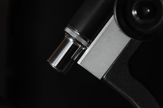
What is meant by a parfocal microscope? Parfocal means that the microscope is binocular. … Parfocal means that when one objective lens is in focus, then the other objectives will also be in focus.
What is parfocal microscope quizlet? Parfocal: A parfocal lens is a microscope that stays approximately in focus when the magnification is changed. For example, if the focal point of a microscope is changed from a low power objective(10x) to a higher power (40x or 100. x), the object stays in focus.
How can you tell if a microscope is parfocal? To determine if a microscope has parfocal objectives, a slide should be brought into focus using the highest magnification settings. The operator should then switch to an objective with a lower magnification level to check for sharpness of focus on the slide.
Which microscope objective is parfocal? Microscopy. Parfocal microscope objectives stay in focus when magnification is changed; i.e., if the microscope is switched from a lower power objective (e.g., 10×) to a higher power objective (e.g., 40×), the object stays in focus. Most modern bright-field microscopes are parfocal.
What is meant by a parfocal microscope? – Related Questions
Why did anton van leeuwenhoek start using microscopes?
Antonie van Leeuwenhoek used single-lens microscopes, which he made, to make the first observations of bacteria and protozoa. His extensive research on the growth of small animals such as fleas, mussels, and eels helped disprove the theory of spontaneous generation of life.
Can chromosomes be seen under light microscope?
Chromosomes, composed of protein and DNA, are distinct dense bodies found in the nucleus of cells. During most of the cell cycle, interphase, the chromosomes are somewhat less condensed and are not visible as individual objects under the light microscope. …
What is the highest magnification your microscope can achieve?
Light microscopes combine the magnification of the eyepiece and an objective lens. Calculate the magnification by multiplying the eyepiece magnification (usually 10x) by the objective magnification (usually 4x, 10x or 40x). The maximum useful magnification of a light microscope is 1,500x.
Can bacteria be seen with a microscope?
Bacteria are too small to see without the aid of a microscope. While some eucaryotes, such as protozoa, algae and yeast, can be seen at magnifications of 200X-400X, most bacteria can only be seen with 1000X magnification. This requires a 100X oil immersion objective and 10X eyepieces..
Can we see microscopic thing in the naked eye?
Experts believe that the naked eye — a normal eye with regular vision and unaided by any other tools — can see objects as small as about 0.1 millimeters. … Until recently, standard microscopes would allow you to see things as small as one micrometer, which is equal to 0.001 mm.
How to clean bacteria off of your microscope?
B) Hydrogen Peroxide can be used with 3% concentration to kill vegetative bacteria, viruses, and fungi. The contact time with surfaces should be at least 10 minutes. Afterwards, rinse with distilled water and let the instrument dry.
What is a microscopic water bear?
Tardigrades are microscopic eight-legged animals that have been to outer space and would likely survive the apocalypse. Bonus: They look like adorable miniature bears. … Like those insects, tardigrades have to shed their cuticles in order to grow.
How to choose which microscope to use?
When Choosing the most important lens in a microscope is the one closest to the specimen. Compound microscopes generally have three, four or five objective lenses, so you can select different magnification levels. The higher the number, or power, of an objective lens, the finer the detail.
When would you use a tem microscope?
The transmission electron microscope is used to view thin specimens (tissue sections, molecules, etc) through which electrons can pass generating a projection image.
What level of magnification was the first microscope capable of?
A Middelburg museum has one of the earliest Janssen microscopes, dated to 1595. It had three sliding tubes for different lenses, no tripod and was capable of magnifying three to nine times the true size. News about the microscopes spread quickly across Europe.
Can you use electron microscope?
Electron microscopy (EM) is a technique for obtaining high resolution images of biological and non-biological specimens. It is used in biomedical research to investigate the detailed structure of tissues, cells, organelles and macromolecular complexes.
What microscope do you need to see tardigrades?
Some notes on equipment: All your really need to find a tardigrade is a microscope, a dish, some water, and time. A small dissecting microscope with a 2-5X objective and 10X eye piece(s) should work fine providing 20-50X magnification.
How to describe a transmission electron microscope?
Transmission electron microscopes (TEM) are microscopes that use a particle beam of electrons to visualize specimens and generate a highly-magnified image. TEMs can magnify objects up to 2 million times. In order to get a better idea of just how small that is, think of how small a cell is.
Can baby aspirin cause microscopic blood in urine?
Certain medications including aspirin, heparin or warfarin (a blood thinner) and anti-cancer drug cyclophosphamide have been known to cause blood in urine. Sometimes, it’s just tiny traces that can only be seen under a microscope. This is called microscopic hematuria.
What is high power on a microscope?
A high-power field (HPF), when used in relation to microscopy, references the field of view under the maximum magnification power of the objective being used. Often, this represents a 400-fold magnification when referenced in scientific papers.
What does light microscope work?
The light microscope is an instrument for visualizing fine detail of an object. It does this by creating a magnified image through the use of a series of glass lenses, which first focus a beam of light onto or through an object, and convex objective lenses to enlarge the image formed.
How to count bacteria microscope?
Abstract: The common practice of counting bacteria using epifluorescence microscopy involves selecting 5–30 random fields of view on a glass slide to calculate the arithmetic mean which is then used to estimate the total bacterial abundance.
Why do microscopes need resolution?
The resolving power of a microscope is the most important feature of the optical system and influences the ability to distinguish between fine details of a particular specimen.
How does a microscope work bbc?
A light microscope uses a series of lenses to produce a magnified image of an object: the object is placed on a rectangular glass slide. … light shines through the object and into the objective lens. the light passes through the eyepiece lens and from there into your eye.
What controls the amount of light on a microscope?
Iris Diaphragm controls the amount of light reaching the specimen. It is located above the condenser and below the stage. Most high quality microscopes include an Abbe condenser with an iris diaphragm. Combined, they control both the focus and quantity of light applied to the specimen.
How to focus a specimen using the compound light microscope?
Look at the objective lens (3) and the stage from the side and turn the focus knob (4) so the stage moves upward. Move it up as far as it will go without letting the objective touch the coverslip. Look through the eyepiece (1) and move the focus knob until the image comes into focus.

