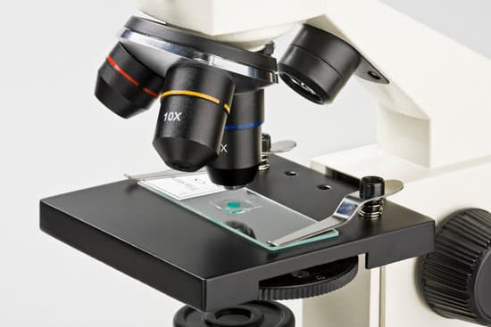
What is meant by parfocal in microscope? : having corresponding focal points all in the same plane : having sets of objectives or eyepieces so mounted that they may be interchanged without varying the focus of the instrument (as a microscope) with which they are used. Other Words from parfocal.
What is parfocal and Parcentral in microscope? A parfocal lens is a microscope that stays approximately in focus when the magnification is changed. For example, if the focal point of a microscope is changed from a low power objective(10x) to a higher power (40x or 100. x), the object stays in focus. Parcentral: The image will remain centered.
What is meant by Par Central and parfocal? Parcentric and parfocal calibration compensate for the deviations from parfocality (focal plane) and parcentricity (collimation) that are normally encountered between different microscope objective lenses. They are both critical for maintaining proper position when changing magnification.
What do we mean by parfocal and resolving power? Parfocal: the objective lenses are mounted on the microscope so that they can be interchanged without having to appreciably vary the focus. Resolving power or resolution: the ability to distinguish objects that are close together. … Magnification: the process of enlarging the size of an object, as an optical image.
What is meant by parfocal in microscope? – Related Questions
What are simple microscope?
A simple microscope is a magnifying glass that has a double convex lens with a short focal length. The examples of this kind of instrument include the hand lens and reading lens. When an object is kept near the lens, then its principal focus with an image is produced, which is erect and bigger than the original object.
Who created the first microscope lens?
Grinding glass to use for spectacles and magnifying glasses was commonplace during the 13th century. In the late 16th century several Dutch lens makers designed devices that magnified objects, but in 1609 Galileo Galilei perfected the first device known as a microscope.
What is the prefix of microscope?
An easy way to remember that the prefix micro- means “small” is through the word microscope, an instrument which allows the viewer to see “small” living things.
Where is the slide containing the specimen placed in microscope?
Since high power microscopes have a very narrow region within which they focus, the object to be viewed (“specimen”) is typically placed on the middle of the slide with another, much thinner square (or circle or rectangle) of glass placed over the specimen.
How to enhance resolving power of microscope?
One way of increasing the optical resolving power of the microscope is to use immersion liquids between the front lens of the objective and the cover slip. Most objectives in the magnification range between 60x and 100x (and higher) are designed for use with immersion oil.
Which microscope uses regular light for illumination?
What Is Fluorescent Microscopy? A fluorescence microscope is much the same as a conventional light microscope with added features to enhance its capabilities. The conventional microscope uses visible light (400-700 nanometers) to illuminate and produce a magnified image of a sample.
What power microscope to see protozoa?
The best type of microscope to use for observation of protozoa is a compound microscope with 3 powers (10x, 40x and 400x). You can use depression slides at the two lower powers but must use a plain slide and coverslip at 400x as the objective will be very close to the specimen when in focus.
Does microscopic hematuria mean bladder cancer?
However, blood in the urine does not necessarily mean a diagnosis of bladder cancer. Infections, kidney stones as well as aspirin and other blood-thinning medications may cause bleeding. In fact, the overwhelming majority of patients who have microscopic hematuria do not have cancer.
Are telescopes and microscopes opposite technology?
Although both instruments magnify objects so that the human eye can see them, a microscope looks at things very near, while telescopes view things very far away. This difference in purpose explains the substantial differences in their design.
What is the microscope used in a school lab?
The most common types of microscopes used in teaching are monocular light microscopes (80%), followed by binocular optical microscopes (16%), digital microscopes (3%), and stereomicroscopes (1%). A total of 43% of teachers perform microscopy using the demonstration method, and 37% of teachers use practical work.
Who invented the most powerful microscope?
Lawrence Berkeley National Labs just turned on a $27 million electron microscope. Its ability to make images to a resolution of half the width of a hydrogen atom makes it the most powerful microscope in the world.
How to count platelets in microscope?
An estimated platelet count can be done quickly with a blood smear evaluated at 100× magnification. At this magnification, the platelet count/µL of blood can be approximated by multiplying the number of platelets seen in one microscope field by 15,000.
How to calculate magnification microscope?
To figure the total magnification of an image that you are viewing through the microscope is really quite simple. To get the total magnification take the power of the objective (4X, 10X, 40x) and multiply by the power of the eyepiece, usually 10X.
What is the microscopic composition and structure of bone?
The basic microscopic unit of bone is an osteon (or Haversian system). Osteons are roughly cylindrical structures that can measure several millimeters long and around 0.2 mm in diameter. Each osteon consists of a lamellae of compact bone tissue that surround a central canal (Haversian canal).
Who invented the usb computer microscope?
An early digital microscope was made by a lens company in Tokyo, Japan in 1986, which is now known as Hirox Co Ltd. It included a control box and a lens connected to a computer. Other versions of digital microscope were later developed by Keyence Corp and Leica Microsystems.
How has the microscope improved over time?
Microscopes became more stable and smaller. Lens improvements solved many of the optical problems that were common in earlier versions. The history of the microscope widens and expands from this point with people from around the world working on similar upgrades and lens technology at the same time.
What can you view with a light microscope?
Explanation: You can see most bacteria and some organelles like mitochondria plus the human egg. You can not see the very smallest bacteria, viruses, macromolecules, ribosomes, proteins, and of course atoms.
What domains include microscopic protists?
All life can be classified into three domains: Bacteria, Archaea, and Eukarya. Organisms in the domain Eukarya keep their genetic material in a nucleus and include the plants, animals, fungi, and protists.
How to find total magnification of a dissecting microscope?
Multiply the magnification on the eyepiece (10x) by any magnification present on the nose piece (usually 1x, but it can be more) by the number on the magnification knob to get your total magnification.
How do you find the magnification of a light microscope?
In order to ascertain the total magnification when viewing an image with a compound light microscope, take the power of the objective lens which is at 4x, 10x or 40x and multiply it by the power of the eyepiece which is typically 10x.
What kind of image do scanning electron microscopes produce?
A scanning electron microscope (SEM) is a type of microscope which uses a focused beam of electrons to scan a surface of a sample to create a high resolution image. SEM produces images that can show information on a material’s surface composition and topography.

