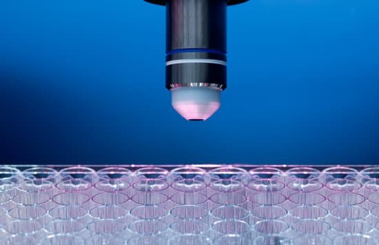
What is methylene blue used for in microscopic studies? Methylene blue is used to stain blood films/smears used in cytology and to stain RNA or DNA for viewing under the microscope or on hybridization membranes. Methylene Blue solution has been used to stain human amniotic fluid stem cells to determine cell viability.
What is the purpose for using methylene blue? As a dye, methylene blue is very useful in the laboratory as an indicator of chemical change and as a biological stain for bacterial cells that are examined under the microscope. The dye highlights the contrasts, revealing details of the cells.
What does methylene blue detect? The methylene blue test is a test to determine the type or to treat methemoglobinemia , a blood disorder.
How do you use methylene blue on a microscope? Microorganisms stain dark blue, whereas proteinaceous material stain light blue. Much less time is required to detect bacteria with Methylene Blue staining than with Gram staining because of this contrast.
What is methylene blue used for in microscopic studies? – Related Questions
Why are microscopes useful tools in biology?
Explanation: The microscope is important because biology mainly deals with the study of cells (and their contents), genes, and all organisms. … Without the microscope, biology would not have been so developed and many diseases would still have no cure.
Why is microscopic phytoplankton very important as a food source?
They provide the base for the entire marine food web. … Zooplankton and other small marine creatures eat phytoplankton and then become food for fish, crustaceans, and other larger species. Phytoplankton make their energy through photosynthesis, the process of using chlorophyll and sunlight to create energy.
Who saw the first bacteria through a microscope?
Antonie van Leeuwenhoek used single-lens microscopes, which he made, to make the first observations of bacteria and protozoa.
Is there an android app that works with a microscope?
Hailed as the best android microscope app for 2020 is the Magnifier and Microscopes, which also works on iPhones and iPads. It’s a really versatile application that can totally transform your phone camera into a magnifier, microscope, macro camera, flashlight, and more.
What is the limit of resolution on a light microscope?
The resolution of the light microscope cannot be small than the half of the wavelength of the visible light, which is 0.4-0.7 µm. When we can see green light (0.5 µm), the objects which are, at most, about 0.2 µm.
How is a comparison microscope used?
A comparison microscope is a device used to observe side-by-side specimens. It consists of two microscopes connected to an optical bridge, which results in a split view window. The comparison microscope is used in forensic sciences to compare microscopic patterns and identify or deny their common origin.
Can we see an electron with an electron microscope?
According to one of the studies in Vienna University of Technology, researchers working on energy-filtered transmission electron microscopy (EFTEM) found out that under given conditions, it is actually possible to view images of individual electrons in their orbit.
What is the microscope field?
Microscope field of view (FOV) is the maximum area visible when looking through the microscope eyepiece (eyepiece FOV) or scientific camera (camera FOV), usually quoted as a diameter measurement (Figure 1).
What does light microscopes mean in biology?
A light microscope is a biology laboratory instrument or tool, that uses visible light to detect and magnify very small objects, and enlarging them. They use lenses to focus light on the specimen, magnifying it thus producing an image.
What is a light microscope formed by?
The light microscope is an instrument for visualizing fine detail of an object. It does this by creating a magnified image through the use of a series of glass lenses, which first focus a beam of light onto or through an object, and convex objective lenses to enlarge the image formed.
What kind of microscope do you view bacteria?
In order to actually see bacteria swimming, you’ll need a lens with at least a 400x magnification. A 1000x magnification can show bacteria in stunning detail.
Why do some scientists use microscopes?
Scientists use microscopes to observe objects too small to view with the human eye. Microscopes can magnify an image hundreds of times while…
What year was the compound microscope invented?
The first compound microscopes date to 1590, but it was the Dutch Antony Van Leeuwenhoek in the mid-seventeenth century who first used them to make discoveries. When the microscope was first invented, it was a novelty item.
Which microscope can magnify objects hundreds of thousands of times?
This makes electron microscopes more powerful than light microscopes. A light microscope can magnify things up to 2000x, but an electron microscope can magnify between 1 and 50 million times depending on which type you use! To see the results, look at the image below.
What do red blood cells look like under a microscope?
Red blood cells are shaped kind of like donuts that didn’t quite get their hole formed. They’re biconcave discs, a shape that allows them to squeeze through small capillaries. This also provides a high surface area to volume ratio, allowing gases to diffuse effectively in and out of them.
When was the first microscope built?
Lens Crafters Circa 1590: Invention of the Microscope. Every major field of science has benefited from the use of some form of microscope, an invention that dates back to the late 16th century and a modest Dutch eyeglass maker named Zacharias Janssen.
Which type of plant is sporophyte dominant with a microscopic?
In the most primitive plants, like mosses, the gametophyte is dominant (i.e. it’s big and green). In higher plants like ferns and fern allies, the sporophyte stage is dominant.
How do you find out the magnification of a microscope?
It’s very easy to figure out the magnification of your microscope. Simply multiply the magnification of the eyepiece by the magnification of the objective lens. The magnification of both microscope eyepieces and objectives is almost always engraved on the barrel (objective) or top (eyepiece).
What is the scientific value of a microscope?
The invention of the microscope allowed scientists to see cells, bacteria, and many other structures that are too small to be seen with the unaided eye. It gave them a direct view into the unseen world of the extremely tiny.
Can inverted telescope work as microscope?
Answer: No, for a telescope, the difference in focal length of the objective and eyepiece is large while in a microscope, the difference of focal lengths is smaller. Therefore, an inverted telescope cannot work as a microscope.
What microscope can see fluorescence?
Most fluorescence microscopes in use are epifluorescence microscopes, where excitation of the fluorophore and detection of the fluorescence are done through the same light path (i.e. through the objective).

