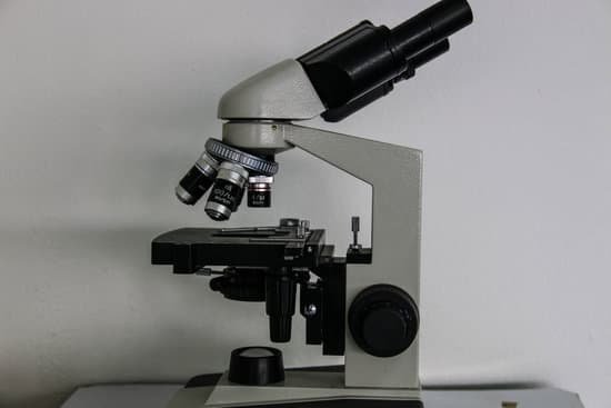
What is needed to see the specimen on a microscope? A microscope is an instrument that can be used to observe small objects, even cells. The image of an object is magnified through at least one lens in the microscope. This lens bends light toward the eye and makes an object appear larger than it actually is.
What does the microscope use to allow you to see the specimen? Light microscopy uses lenses to focus light on a specimen to produce an image. Commonly used light microscopes include brightfield, darkfield, phase-contrast, differential interference contrast, fluorescence, confocal, and two-photon microscopes.
How do you view a specimen under a microscope? Scan the slide (right to left and top to bottom) at low power to get an overview of the specimen. Then center the part of the specimen you want to view at higher power. Rotate the nosepiece to the 10x objective for 100x magnification. Refocus and view your specimen carefully.
What is used to view a specimen? Microscopes are designated as either light microscopes or electron microscopes. The former use visible light or ultraviolet rays to illuminate specimens. They include brightfield, darkfield, phase-contrast, and fluorescent instruments.
What is needed to see the specimen on a microscope? – Related Questions
When to use confocal microscope?
As a distinctive feature, confocal microscopy enables the creation of sharp images of the exact plane of focus, without any disturbing fluorescent light from the background or other regions of the specimen. Therefore, structures within thicker objects can be conveniently visualized using confocal microscopy.
What is a microscope stage in science?
microscope stage – a small platform on a microscope where the specimen is mounted for examination. stage. platform – a raised horizontal surface; “the speaker mounted the platform”
What is a simple microscope used to look at?
A simple microscope is used to see the magnified image of an object. Antonie Van Leeuwenhoek, a Dutch, invented the first simple microscope, consisting of a small single high powered converging lens to inspect the small micro-organisms of freshwater.
How are microscopic transistors made?
In production, transistors are “printed” on a silicon wafer through a complex process called lithography. To produce the 7 nm chip, the team employed a new type of lithography in the manufacturing process, Extreme Ultraviolet, or EUV, which delivers huge improvements over today’s mainstream optical lithography.
Who invented the scanning helium ion microscope?
Notte and ALIS colleagues Bill Ward and Nick Economou developed the helium gas ion source, which is the HeIM’s distinguishing component. Zeiss acquired ALIS and then developed the HeIM, which is designed to collect images via two modes: secondary-electron mode and RBI mode.
How to measure magnification of a microscope?
It’s very easy to figure out the magnification of your microscope. Simply multiply the magnification of the eyepiece by the magnification of the objective lens. The magnification of both microscope eyepieces and objectives is almost always engraved on the barrel (objective) or top (eyepiece).
How does robert hooke microscope work?
To combat dark specimen images, Hooke designed an ingenious method of concentrating light on his specimens, as shown in the illustration. He passed light generated from an oil lamp through a water-filled glass flask to diffuse the light and provide a more even and intense illumination for the samples.
How electron microscope differs from light microscopy?
Electron microscopes differ from light microscopes in that they produce an image of a specimen by using a beam of electrons rather than a beam of light. Electrons have much a shorter wavelength than visible light, and this allows electron microscopes to produce higher-resolution images than standard light microscopes.
What year was the transmission electron microscope invented?
Ernst Ruska at the University of Berlin, along with Max Knoll, combined these characteristics and built the first transmission electron microscope (TEM) in 1931, for which Ruska was awarded the Nobel Prize for Physics in 1986.
How is an electron microscope similar to a light microscope?
Light Microscope vs Electron Microscope. Light microscopes and electron microscopes both use radiation – in the form of either light or electron beams, to form larger and more detailed images of objects (e.g. biological specimens, materials, crystal structures, etc.) than the human eye can produce unaided.
What is a good magnification for a microscope?
Lower magnification (10-20x) produces a larger field of view and is best for young kids. It is also ideal for viewing stamps and coins. Higher magnification (30-40x) is better for close-ups and more detailed work.
What is a raster microscope?
The term raster, in laser raster microscopy, refers to the linear direction of the laser beam’s motion, which is controlled either by highly-tunable dichroic mirrors inside a fluorescence microscope, or by minutely moving the sample.
How to determine the magnification power of a microscope?
To figure the total magnification of an image that you are viewing through the microscope is really quite simple. To get the total magnification take the power of the objective (4X, 10X, 40x) and multiply by the power of the eyepiece, usually 10X.
What is depth of focus microscope?
The “focal depth (depth of focus)” is the range of distances for which the object is imaged with an acceptable sharpness on the image plane. The focal depth is proportional to the spatial resolution of a microscope and to the square of magnification, and inversely proportional to the aperture angle.
How to calculate total magnification on a microscope?
To figure the total magnification of an image that you are viewing through the microscope is really quite simple. To get the total magnification take the power of the objective (4X, 10X, 40x) and multiply by the power of the eyepiece, usually 10X.
How do laser scanning confocal microscopes work?
A confocal microscope works with a laser and pinhole spatial filters. The laser provides the excitation light, and the laser light reflects off a mirror. … The mirrors scan the laser across the sample. The dye and the emitted light get descanned by the mirrors that scan the excitation light.
Which kinds of lenses are in a light microscope quizlet?
A compound light microscope uses two lenses at the same time to view objects-the objective lens, which gathers light and magnifies the image of the object, and the ocular lens, which one looks through and which further magnifies the image.
Who made the dissecting microscope?
It was first designed by Cherudin d’Orleans in 1677 by making a small microscope with two separate eyepieces and objective lenses.
Do light microscopes have better resolution than?
Electron microscopes differ from light microscopes in that they produce an image of a specimen by using a beam of electrons rather than a beam of light. Electrons have much a shorter wavelength than visible light, and this allows electron microscopes to produce higher-resolution images than standard light microscopes.
Can scanning electron microscope view living specimens?
Electron microscopes use a beam of electrons instead of beams or rays of light. Living cells cannot be observed using an electron microscope because samples are placed in a vacuum. … the scanning electron microscope (SEM) has a large depth of field so can be used to examine the surface structure of specimens.
What is the microscopic morphology?
Morphology refers to the size, shape, and arrangement of cells. The observation of microbial cells requires not only the use of microscopes but also the preparation of the cells in a manner appropriate for the particular kind of microscopy.

