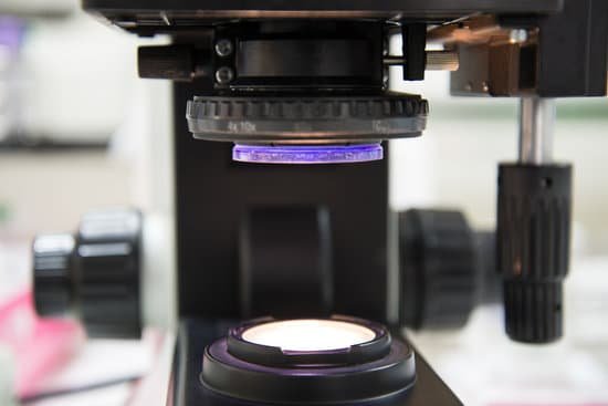
What is reflected light used for in a microscope? Reflected light microscopy is primarily used to examine opaque specimens that are inaccessible to conventional transmitted light techniques.
How is reflection used in microscopes? In reflected light microscopy, illuminating light reaches the specimen, which may absorb some of the light and reflect some of the light, either in a specular or diffuse manner. Light that is returned upward can be captured by the objective in accordance with the objective’s numerical aperture.
What do light microscopes use to reflect light? The reflected light microscope use ingenious systems of mirrors, prisms and semi-mirrored glasses which let pass the light in one direction and reflects in the other. Polarizing microscopy can be used with reflected and transmitted light.
What is the light used for in a microscope? A light microscope is an optical instrument used to view objects too small to with the naked eye. It is so-called because it employs the use of white or visible light to illuminate the object of interest so it can be magnified and viewed through one or a series of lenses.
What is reflected light used for in a microscope? – Related Questions
Do light microscope lens form real or virtual images?
The objective lens is positioned close to the object to be viewed. It forms an upside-down and magnified image called a real image because the light rays actually pass through the place where the image lies. … Because light rays do not actually pass through this location, the image is called a virtual image.
Where is condenser iris in a microscope?
Iris Diaphragm controls the amount of light reaching the specimen. It is located above the condenser and below the stage. Most high quality microscopes include an Abbe condenser with an iris diaphragm.
How does a compound microscope differ from a simple microscope?
A magnifying instrument that uses two types of lens to magnify an object with different zoom levels of magnification is called a compound microscope. … A magnifying instrument that uses only one lens to magnify objects is called a Simple microscope.
What magnification to hooke’s microscope?
Which was better? Some of Leeuwenhoek’s simple microscopes could magnify objects more than 250 times, but Hooke’s compound microscopes only magnified somewhere between 20 and 50 times.
How to describe simple cuboidal epithelium under a microscope?
A cuboidal epithelial cell looks close to a square. A columnar epithelial cell looks like a column or a tall rectangle. A few epithelial layers are constructed from cells that are said to have a transitional shape.
Which microscope is used to visualize live cells?
The cells are visualized using a confocal microscope equipped for live imaging or a spinning disc microscope. Fluorescent microscopes can also be used. To reduce the movement of the cells, they can be plated on concanavalin A-coated coverslips.
How to see sperm in microscope?
You can view sperm at 400x magnification. You do NOT want a microscope that advertises anything above 1000x, it is just empty magnification and is unnecessary. In order to examine semen with the microscope you will need depression slides, cover slips, and a biological microscope.
How do you raise a stage on a microscope?
Turn the revolving turret (2) so that the lowest power objective lens (eg. 4x) is clicked into position. Place the microscope slide on the stage (6) and fasten it with the stage clips. Look at the objective lens (3) and the stage from the side and turn the focus knob (4) so the stage moves upward.
Do you have to use a microscope to see lice?
A visual inspection of your hair and scalp is usually effective in detecting head lice, though the creatures are so small that they can be difficult to spot with the naked eye.
Which part of the microscope is the most important?
While the modern microscope has many parts, the most important pieces are its lenses. It is through the microscope’s lenses that the image of an object can be magnified and observed in detail.
What do telescopes microscopes and cameras use?
Optical instruments such as microscopes, telescopes, and cameras use mirrors and lenses to reflect and refract light and form images.
How do chemical stains make light microscope?
How do chemical stains make light microscopes more useful? They make it useful because it helps show specific structures in the cell. What are two main types of electron microscopes?
Can you see cells with a light microscope?
Since most cells are between 1 and 100 μm in diameter, they can be observed by light microscopy, as can some of the larger subcellular organelles, such as nuclei, chloroplasts, and mitochondria.
What does a animal cell look like under a microscope?
Under the microscope, animal cells appear different based on the type of the cell. However, the internal structure and organelles are more or less similar. Animal cells usually are transparent and colorless, and the thickness of the cell differs throughout the cytoplasm.
Why is microscope called compound?
The common light microscope used in the laboratory is called a compound microscope because it contains two types of lenses that function to magnify an object.
How to view a specimen on a microscope?
Place your sample on the stage (3) and turn on the LED light (2). Look through the eyepieces (4) and move the focus knob (1) until the image comes into focus. Adjust the distance between the eyepieces (4) until you can see the sample clearly with both eyes simultaneously (you should see the sample in 3D).
Can you see probiotic with microscope?
Dried probiotics can indeed be shipped in the summer heat, with very little mortality. Bacteria can indeed be observed and counted with an inexpensive microscope. … For viewing bacteria, a probiotic is nice and safe to handle.
Where is the fine focus adjustment knob on a microscope?
Fine Adjustment Knob – This knob is inside the coarse adjustment knob and is used to bring the specimen into sharp focus under low power and is used for all focusing when using high power lenses.
Why is the typical lab microscope called a compound microscope?
The common light microscope used in the laboratory is called a compound microscope because it contains two types of lenses that function to magnify an object.
How does yeast look like under a microscope?
When viewing the specimen under high magnification (1000x and above) one will see oval (egg shaped) organism, which are the yeast. It is also possible to observe the buds, which can be seen on some of the yeast cells.
Can distinguish mitochondria under a light microscope?
Mitochondria are visible with the light microscope but can’t be seen in detail. Ribosomes are only visible with the electron microscope.

