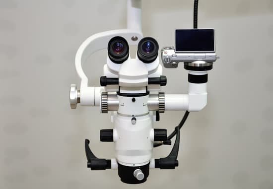
What is the advantage of using transmission electron microscope? The advantage of the transmission electron microscope is that it magnifies specimens to a much higher degree than an optical microscope. Magnification of 10,000 times or more is possible, which allows scientists to see extremely small structures.
What are the advantages of a transmission electron microscope? A Transmission Electron Microscope produces images via the interaction of electrons with a sample.
What are the advantages and disadvantages of a transmission electron microscope? iv) TEMs provide the highest magnification in microscope field. v) TEMs can provide information about surface features, shape, size and structure. However, TEMs also present some disadvantages: i) The instruments are very large and expensive.
What is one advantage of a scanning electron microscope over a transmission electron microscope? Pros. The power of SEM cannot be underestimated. The process by which the focused beam of electrons creates a magnified image is so advanced that the magnification is anywhere between 10 and 1,000,000 times. As such, it is a key tool for basic research, as well as quality control and failure analysis.
What is the advantage of using transmission electron microscope? – Related Questions
What cells can be seen with an electron microscope?
The cell wall, nucleus, vacuoles, mitochondria, endoplasmic reticulum, Golgi apparatus, and ribosomes are easily visible in this transmission electron micrograph.
Can sandstone have microscopic quartz grains?
Grains can include quartz or chert rock fragments. Quartz arenites are texturally mature to supermature sandstones. These pure quartz sands result from extensive weathering that occurred before and during transport. This weathering removed everything but quartz grains, the most stable mineral.
How many lenses did robert hooke’s microscope have?
Hooke’s microscope was a much larger, ‘compound’ instrument. It used three lenses: a small double-convex eye-lens at the top, then a large plano-convex field-lens, and another double-convex lens with a short focal length at the bottom of the tube.
What can be used to preserve microscope specimen?
To keep your prepared microscope slides in good condition, always store them in a container made for the purpose and away from heat and bright light. The ideal storage area is a cool, dark location, such as a closed cabinet in a temperature-controlled room. Stained slides naturally fade over time.
How are comparison microscopes used to analyze fired bullets?
These patterns or imperfections, therefore, amount to a “signature” that each barrel imprints on each of the bullets fired through it. … Comparison microscope is used to analyze the matching of the microscopic impressions found on the surface of bullets and casings.
How much is a compound microscope?
The most popular compound microscopes from some of the most well-known brands cost on average around $900-$1,200, although there are beginner microscopes that are just above the toy level that cost $100.
What is a microscope slide cover used for in biology?
When viewing any slide with a microscope, a small square or circle of thin glass called a coverslip is placed over the specimen. It protects the microscope and prevents the slide from drying out when it’s being examined. The coverslip is lowered gently onto the specimen using a mounted needle .
How to tell bacteria through microscope?
In order to see bacteria, you will need to view them under the magnification of a microscopes as bacteria are too small to be observed by the naked eye. Most bacteria are 0.2 um in diameter and 2-8 um in length with a number of shapes, ranging from spheres to rods and spirals.
What is a compound light microscope used for in biology?
Compound microscopes are used to view small samples that can not be identified with the naked eye. These samples are typically placed on a slide under the microscope.
Are there any microscopic bugs?
Some biting bugs are so tiny that even bed bugs, fleas, and ticks are more prominent than them. These bugs are known as microscopic bugs. Some of these microscopic bugs are parasites on humans. And some are outdoor bugs that sneak inside your home to bite you and hide inside your home.
Is there a microscope stronger than an electron one?
The first of two advanced microscopes has been installed at Lawrence Berkeley National Laboratory. TEAM 0.5 is the world’s most powerful transmission electron microscope and is capable of producing images with half-angstrom resolution, less than the diameter of a single hydrogen atom.
Where should the focal lens be when making a microscope?
1. To build your microscope, place the lens identified as the eyepiece (ocular) lens on the end of the cardboard tube having the smallest diameter. 2. Take the other lens, the one identified as the objective lens, and place it on the end of the cardboard tube having the largest diameter.
What did louis pasteur discover by using a compound microscope?
Pasteur discovered another compound in wine called paratartaric acid that had the same chemical composition as tartaric acid. Most scientists assumed the two substances were the same.
How to find resolving power of microscope physics?
(1.22λ)/ (2n sinβ) is known as numerical aperture of the objective lens. Resolving Power(R.P) of a microscope ∝ (1/dmin). This implies resolving power decreases as the distance increases.
What type of microscope is used to examine hairs?
A stereo microscope is typically used for the initial examination of hair (mounted and unmounted) before moving on to the compound microscope.
Why do objects appear upside down in a microscope?
Microscopes invert images which makes the picture appear to be upside down. The reason this happens is that microscopes use two lenses to help magnify the image. Some microscopes have additional magnification settings which will turn the image right-side-up.
What is the advantage of electron microscope over light microscope?
Electron microscopes have two key advantages when compared to light microscopes: They have a much higher range of magnification (can detect smaller structures) They have a much higher resolution (can provide clearer and more detailed images)
How does a quantum microscope work?
They used a type of microscope with two laser light sources, but sent one of the beams through a specially designed crystal that “squeezes” the light. It does so by introducing quantum correlations in the photons – the particles of light in the laser beam.
What is a fiber optic microscope used for?
A Fiber Microscope is used for inspecting fiber terminations. This CL series utilizes white LED for coaxial illumination and provides the most critical view of ferrule end face. It has good optical performance and integrated safety filters and produces excellent detail of scratches and contamination.
How does a basic 2 lens microscope work?
A compound microscope uses two or more lenses to produce a magnified image of an object, known as a specimen, placed on a slide (a piece of glass) at the base. … By raising and lowering the stage, you move the lenses closer to or further away from the object you’re examining, adjusting the focus of the image you see.
What microscope can produce three dimensional images of cells?
Scanning electron microscopy (SEM) is a powerful technique, traditionally used for imaging the surface of cells, tissues and whole multicellular organisms (see An Introduction to Electron Microscopy for Biologists)(Fig. 1).

