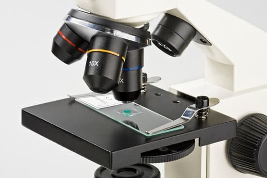
What is the definition of microscope slide in science? microscope slide Add to list Share. Definitions of microscope slide. a small flat rectangular piece of glass on which specimens can be mounted for microscopic study.
What is a microscope slide in science? A microscope slide is a thin flat piece of glass, typically 75 by 26 mm (3 by 1 inches) and about 1 mm thick, used to hold objects for examination under a microscope. Typically the object is mounted (secured) on the slide, and then both are inserted together in the microscope for viewing.
What do you call a microscope slide? A glass slide is a thin, flat, rectangular piece of glass that is used as a platform for microscopic specimen observation. A typical glass slide usually measures 25 mm wide by 75 mm, or 1 inch by 3 inches long, and is designed to fit under the stage clips on a microscope stage.
What is a slide science? A microscope slide is a thin sheet of glass used to hold objects for examination under a microscope. … The cover glass serves two purposes: (1) it protects the microscope’s objective lens from contacting the specimen, and (2) it creates an even thickness (in wet mounts) for viewing.
What is the definition of microscope slide in science? – Related Questions
What is meant by compound light microscope?
A compound light microscope is a microscope with more than one lens and its own light source. In this type of microscope, there are ocular lenses in the binocular eyepieces and objective lenses in a rotating nosepiece closer to the specimen.
Who discovered transmission electron microscope?
Ernst Ruska at the University of Berlin, along with Max Knoll, combined these characteristics and built the first transmission electron microscope (TEM) in 1931, for which Ruska was awarded the Nobel Prize for Physics in 1986.
What is macroscopic and microscopic approach?
Microscopic approach considers the behaviour of every molecule by using statistical methods. In Macroscopic approach we are concerned with the gross or average effects of many molecules’ infractions. These effects, such as pressure and temperature, can be perceived by our senses and can be measured with instruments.
Can you see dna during interphase under microscope?
Prokaryotic chromosomes are less condensed than their eukaryotic counterparts and don’t have easily identified features when viewed under a light microscope. Figure 2: A the appearance of DNA during interphase versus mitosis. During interphase, the cell’s DNA is not condensed and is loosely distributed.
Where is the coarse adjustment knob on a microscope?
Coarse Adjustment Knob- The coarse adjustment knob located on the arm of the microscope moves the stage up and down to bring the specimen into focus. The gearing mechanism of the adjustment produces a large vertical movement of the stage with only a partial revolution of the knob.
Are lenses used in microscopes?
A typical microscope, a compound microscope, uses several lenses and a light source to greatly enhance the image of the object you are viewing. The compound microscope uses a system of lenses that work together to increase the size of the image.
What is evos microscope?
The EVOS XL Core system is an integrated transmitted-light inverted imaging system that combines high-quality optics, a 12.1′ high-resolution LCD display, and a digital color camera. … You will be astonished at how easy it is to operate and amazed at how extraordinary your images look on-screen.
Do a light or electron microscopes have higher magnification?
Electron microscopes allow for higher magnification in comparison to a light microscope thus, allowing for visualization of cell internal structures.
What is the magnification of the transmission electron microscope?
Transmission electron microscopes (TEM) are microscopes that use a particle beam of electrons to visualize specimens and generate a highly-magnified image. TEMs can magnify objects up to 2 million times.
What do cells actually look like under a microscope?
Under a low power microscope, the cell membrane is observed as a thin line, while the cytoplasm is completely stained. The cell organelles are seen as tiny dots throughout the cytoplasm, whereas the nucleus is seen as a thick drop.
Where do you place the slide on a microscope?
Place the microscope slide on the stage (6) and fasten it with the stage clips. Look at the objective lens (3) and the stage from the side and turn the focus knob (4) so the stage moves upward. Move it up as far as it will go without letting the objective touch the coverslip.
Can be viewed with light microscope?
Explanation: You can see most bacteria and some organelles like mitochondria plus the human egg. You can not see the very smallest bacteria, viruses, macromolecules, ribosomes, proteins, and of course atoms.
Which color wavelength is used in microscopes?
Under most circumstances, microscopists use white light generated by a tungsten-halogen bulb to illuminate the specimen. The visible light spectrum is centered at about 550 nanometers, the dominant wavelength for green light (our eyes are most sensitive to green light).
How did the invention of the microscope affect scientific theories?
Explanation: With the development and improvement of the light microscope, the theory created by Sir Robert Hooke that organisms would be made of cells was confirmed as scientist were able to actually see cells in tissues placed under the microscope.
What is resolution of microscope?
In microscopy, the term ‘resolution’ is used to describe the ability of a microscope to distinguish detail. In other words, this is the minimum distance at which two distinct points of a specimen can still be seen – either by the observer or the microscope camera – as separate entities.
How to calculate vernier constant of travelling microscope?
We know that the main scale division of a travelling microscope is the smallest division on the main scale. Where, VC is the Vernier constant or basically the least count which is defined as the ratio of the smallest division of the main scale (MSD) divided by the number of divisions on the vernier scale.
What are the similarities between a telescope and a microscope?
Microscopes and telescopes are quite similar in that they are both utilized to view objects up close. The utilization of microscopes and telescopes dates back to the early 17th century and the similarity in the use of convex and concave mirror and lenses to make them have not changed much in the last few centuries.
How to see mites microscope?
Place the slide glass side up on your microscope and look. The mites will be found at the end of the hair follicles. Use between 40x and 100x magnification.
What accidents might happen when using a microscope?
Conclusions: The most common occupational concerns of microscope users were musculoskeletal problems of neck and back regions, eye fatigue, aggravation of ametropia, headache, stress due to long working hours and anxiety during or after microscope use.
How did the simple microscope work?
A simple light microscope manipulates how light enters the eye using a convex lens, where both sides of the lens are curved outwards. When light reflects off of an object being viewed under the microscope and passes through the lens, it bends towards the eye. This makes the object look bigger than it actually is.
Did robert hooke make a microscope?
Although Hooke did not make his own microscopes, he was heavily involved with the overall design and optical characteristics. The microscopes were actually made by London instrument maker Christopher Cock, who enjoyed a great deal of success due to the popularity of this microscope design and Hooke’s book.

