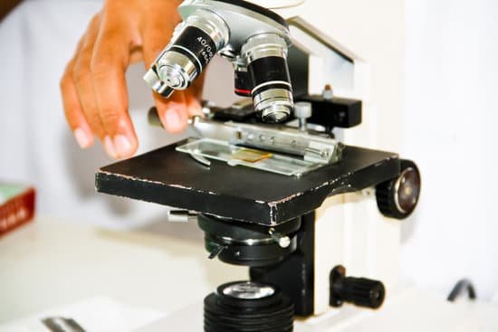
What is the field of view microscope? Introduction. Microscope field of view (FOV) is the maximum area visible when looking through the microscope eyepiece (eyepiece FOV) or scientific camera (camera FOV), usually quoted as a diameter measurement (Figure 1).
How do you find the field of view of a microscope? For instance, if your eyepiece reads 10X/22, and the magnification of your objective lens is 40. First, multiply 10 and 40 to get 400. Then divide 22 by 400 to get a FOV diameter of 0.055 millimeters.
What is the field of view on a microscope 40x?
How do you describe field of view? Field of view (FOV) is the open observable area a person can see through his or her eyes or via an optical device. In the case of optical devices and sensors, FOV describes the angle through which the devices can pick up electromagnetic radiation. FOV allows for coverage of an area rather than a single focused point.
What is the field of view microscope? – Related Questions
What is an objective lens in a microscope?
The objective lens consists of several lenses to magnify an object and project a larger image. According to the difference of the focal distance, lenses of different magnifications are available, such as 4x, 10x, 40x, and 50x.
Can an electron microscope look at live specimens?
Electron microscopes use a beam of electrons instead of beams or rays of light. Living cells cannot be observed using an electron microscope because samples are placed in a vacuum.
What does differential interference contrast microscope produce?
Differential Interference Contrast (DIC) is a microscopy technique that introduces contrast to images of specimens which have little or no contrast when viewed using brightfield microscopy. … Using DIC produces high resolution images with good contrast. It is best for visualising unstained samples.
How to convert a microscope to digital?
One of the simplest methods is to mount a microscope camera over the microscope eyepiece with an Over-Eyepiece Adapter. A microscope camera has a USB connection that plugs directly into your computer. When the software is opened you can view a live image from the microscope on the computer.
What does the ocular do on a microscope?
The eyepiece, or ocular lens, is the part of the microscope that magnifies the image produced by the microscope’s objective so that it can be seen by the human eye.
Do dissecting microscopes invert images?
Compound microscopes invert images! … Quite a few microscopes, including electron microscopes and digital microscopes, will not show you inverted images. Binocular and dissecting microscopes will also not show an inverted image because of their increased level of magnification.
Why are mirrors important to the function of a microscope?
Optical microscopes make extensive use of planar mirrors, both for directing the illumination beam through the optical pathway and onto the specimen, and to project images into the eyepieces or an image sensor.
How do you figure out total magnification on a microscope?
To figure the total magnification of an image that you are viewing through the microscope is really quite simple. To get the total magnification take the power of the objective (4X, 10X, 40x) and multiply by the power of the eyepiece, usually 10X.
Can you view living microbes with electron microscope?
Preparation of a specimen for viewing under an electron microscope will kill it; therefore, live cells cannot be viewed using this type of microscopy. In addition, the electron beam moves best in a vacuum, making it impossible to view living materials.
What is the meaning microscope stage?
Microscope Stages. All microscopes are designed to include a stage where the specimen (usually mounted onto a glass slide) is placed for observation. Stages are often equipped with a mechanical device that holds the specimen slide in place and can smoothly translate the slide back and forth as well as from side to side …
What to eat if you have microscopic colitis?
These include applesauce, bananas, melons and rice. Avoid high-fiber foods such as beans and nuts, and eat only well-cooked vegetables. If you feel as though your symptoms are improving, slowly add high-fiber foods back to your diet. Eat several small meals rather than a few large meals.
What are the parts of microscope and meaning?
Eyepiece Lens: the lens at the top that you look through, usually 10x or 15x power. Tube: Connects the eyepiece to the objective lenses. Arm: Supports the tube and connects it to the base. Base: The bottom of the microscope, used for support. Illuminator: A steady light source (110 volts) used in place of a mirror.
Does a dissecting microscope invert images?
Compound microscopes invert images! … Quite a few microscopes, including electron microscopes and digital microscopes, will not show you inverted images. Binocular and dissecting microscopes will also not show an inverted image because of their increased level of magnification.
How much does a scanning microscope cost?
The cost of a scanning electron microscope (SEM) can range from $80,000 to $2,000,000. The cost of a transmission electron microscope (TEM) can range from $300,000 to $10,000,000. The cost of a focused ion beam electron microscope (FIB) can range from $500,000 to $4,000,000.
What is the smallest object a light microscope can see?
The smallest thing that we can see with a ‘light’ microscope is about 500 nanometers. A nanometer is one-billionth (that’s 1,000,000,000th) of a meter. So the smallest thing that you can see with a light microscope is about 200 times smaller than the width of a hair. Bacteria are about 1000 nanometers in size.
What is the nosepiece of a microscope used for?
Nosepiece houses the objectives. The objectives are exposed and are mounted on a rotating turret so that different objectives can be conveniently selected. Standard objectives include 4x, 10x, 40x and 100x although different power objectives are available. Coarse and Fine Focus knobs are used to focus the microscope.
How to measure cells under a light microscope?
You can use an ocular micrometer to measure cell size. An ocular micrometer is basically a tiny ruler etched into one of the ocular lenses; it can give you a better estimate of the size of a cell, provided you calibrate it with a stage micrometer, which is a microscope slide that has a scale etched into its surface.
What microscope first saw the atom?
Of the 1955 microscope, Dr. Mueller recalled later: “It was a sticky day in August that I became the first person to see an atom. On that day, the regular array of atoms and a crystal lattice became clearly visible through the field ion microscope which I had developed.”
What does the coarse adjustment knob of a microscope do?
4. COARSE ADJUSTMENT KNOB — A rapid control which allows for quick focusing by moving the objective lens or stage up and down. It is used for initial focusing.
Did the rife microscope work?
The technology he described is now classified as a type of radionics device which has been rejected as an effective medical treatment.
How to estimate actual length of cell under microscope?
Divide the number of cells in view with the diameter of the field of view to figure the estimated length of the cell. If the number of cells is 50 and the diameter you are observing is 5 millimeters in length, then one cell is 0.1 millimeter long. Measured in microns, the cell would be 1,000 microns in length.

