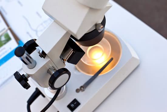
What is the function of mirror in microscope? If your microscope has a mirror, it is used to reflect light from an external light source up through the bottom of the stage.
Why is it important to calibrate each objective? Why is it necessary to calibrate the ocular micrometer with each objective? … The magnification is different for each objective lens. The numerical value holds only for the specific objective-ocular lens combination O.S. changes with each objective change each microscope is different.
What is the purpose of calibrating the ocular micrometer? This is a simple and precise method for measuring objects seen in the microscope. Ocular micrometers are calibrated by comparing the ocular micrometer scale with a calibrated stage micrometer. A calibration procedure must be completed to determine the calibration factor for each objective and each microscope.
What is calibration factor of microscope? number of divisions on stage micrometer divided by the number of divisions on the eyepiece. Example: … Here, the number obtained from the calculation is the calibration factor and gives the number of units in each division of the eyepiece.
What is the function of mirror in microscope? – Related Questions
How should a microscope be carried quizlet?
How should a microscope be carried? It should be carried by one hand under the base and one holding the arm. The ocular and objectives are found at the top and bottom of what part of a microscope? It is the body tube.
How to find total magnification on a microscope?
To figure the total magnification of an image that you are viewing through the microscope is really quite simple. To get the total magnification take the power of the objective (4X, 10X, 40x) and multiply by the power of the eyepiece, usually 10X.
Where is the microscope in sims 4 scientist career?
Life Under a Microscope is a large microscope in The Sims 4. It costs §1,630 and is found in the Activities and Skills section in build mode, under Knowledge.
Which scientist invented the very first optical microscopes?
Zacharias Janssen, credited with inventing the microscope. (Image credit: Public domain.) For millennia, the smallest thing humans could see was about as wide as a human hair. When the microscope was invented around 1590, suddenly we saw a new world of living things in our water, in our food and under our nose.
What is the science of the microscope?
Microscopy is the science of investigating small objects and structures using a microscope. … The most common microscope (and the first to be invented) is the optical microscope, which uses lenses to refract visible light that passed through a thinly sectioned sample to produce an observable image.
What is the ocular on a microscope?
Eyepieces (Oculars) The eyepiece, or ocular lens, is the part of the microscope that magnifies the image produced by the microscope’s objective so that it can be seen by the human eye.
Which microscope can see hair?
A stereo microscope is good for the initial examination of hair before moving on to a compound microscope. Under a stereo microscope, you can easily observe basic characteristics such as color, shape, texture, and length of hair.
Is a glass microscope slide a conductor or insulator?
Glass coated in Indium Tin Oxide (ITO) is conductive and optically transparent, making it suitable for microscopy slides. Glass coated in Indium Tin Oxide (ITO) is conductive and optically transparent, making it highly suitable for use in microscopy slides.
What is the main advantage of the scanning electron microscope?
Advantages of a Scanning Electron Microscope include its wide-array of applications, the detailed three-dimensional and topographical imaging and the versatile information garnered from different detectors.
What is an advantage of using an electron microscope?
Electron microscopes have two key advantages when compared to light microscopes: They have a much higher range of magnification (can detect smaller structures) They have a much higher resolution (can provide clearer and more detailed images)
What is the function of the following microscope parts condenser?
Condenser is used to collect and focus the light from the illuminator on to the specimen. It is located under the stage often in conjunction with an iris diaphragm. Iris Diaphragm controls the amount of light reaching the specimen. It is located above the condenser and below the stage.
What are the two different types of electron microscopes?
Today there are two major types of electron microscopes used in clinical and biomedical research settings: the transmission electron microscope (TEM) and the scanning electron microscope (SEM); sometimes the TEM and SEM are combined in one instrument, the scanning transmission electron microscope (STEM):
Why is the microscope important in the study of zoology?
Explanation: The microscope is important because biology mainly deals with the study of cells (and their contents), genes, and all organisms. Some organisms are so small that they can only be seen by using magnifications of ×2000−×25000 , which can only be achieved by a microscope.
Who developed a compound microscope to observe small organisms?
Zaccharias Janssen, along with his father Hans, may have invented the telescope, the simple microscope, and the compound microscope during the late 1500s or early 1600s.
What is the use of binocular microscope?
A binocular microscope is any optical microscope with two eyepieces to significantly ease viewing and cut down on eye strain.
What kind of oil is used with microscope?
Immersion oils are transparent oils that have specific optical and viscosity characteristics necessary for use in microscopy. Typical oils used have an index of refraction of around 1.515. An oil immersion objective is an objective lens specially designed to be used in this way.
What are the optical parts of the microscope?
The microscope optical train typically consists of an illuminator (including the light source and collector lens), a substage condenser, specimen, objective, eyepiece, and detector, which is either some form of camera or the observer’s eye (Table 1).
What is the diaphragm function in the microscope?
The field diaphragm controls how much light enters the substage condenser and, consequently, the rest of the microscope.
What does 10x 20 mean on microscope?
The eyepieces will generally have an inscription on them such as WF10X/20. This means the eyepieces are 10x magnification with a 20mm field of view. The objective lens value is typically printed on the edge of the objective, or on the zoom knob on the side of the microscope.
What is microscopic collagenous colitis?
Collagenous colitis, in which a thick layer of protein (collagen) develops in colon tissue. Lymphocytic colitis, in which white blood cells (lymphocytes) increase in colon tissue. Incomplete microscopic colitis, in which there are mixed features of collagenous and lymphocytic colitis.

