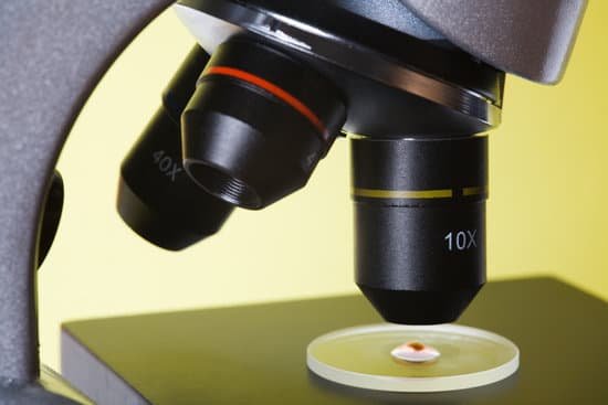
What is the image produced by a microscope? The objective lens is positioned close to the object to be viewed. It forms an upside-down and magnified image called a real image because the light rays actually pass through the place where the image lies.
Can we see DNA through microscope? Yes, but not in detail. “Many scientists use electron, scanning tunneling and atomic force microscopes to view individual DNA molecules,” said Michael W. Davidson, curator of the National High Magnetic Field Laboratory at Florida State University.
What type of microscope can you see DNA? Electron microscopes, too, can see DNA in cells, and DNA sequencers can determine the A’s, T’s, C’s, and G’s (nucleotides) it’s made of.
Can we see our sperm? Can you actually see sperm? Only if you’re looking through a microscope. Sperm are tiny. Like really tiny.
What is the image produced by a microscope? – Related Questions
How long must grid dry before using electron microscope?
Always let the paint fully dry before loading the sample in the microscope. Typically, 30 minutes is long enough for the solvents to evaporate, hence preventing chamber contamination and imaging issues.
What do pinworm eggs look like under a microscope?
The pinworms are white, can be seen with the naked eye (no magnification), and are about the length of a staple (about 8-13 mm for female and 2-5mm for male worms). The eggs that are laid by the female worms are not visible as they are about 55 micrometers in diameter and are translucent (see Figure 1).
What is the main difference between microscopic and macroscopic?
The macroscopic level includes anything seen with the naked eye and the microscopic level includes atoms and molecules, things not seen with the naked eye. Both levels describe matter.
Can you identify fire ants with a microscope?
Identifying the specific species of fire ant is easier if you have access to a microscope and a good light source. Many of the features used to identify fire ants to species are small and hard to see.
How to find total magnification of a compound microscope?
To get the total magnification take the power of the objective (4X, 10X, 40x) and multiply by the power of the eyepiece, usually 10X.
What is an advantage of electron microscopes?
Electron microscopes have two key advantages when compared to light microscopes: They have a much higher range of magnification (can detect smaller structures) They have a much higher resolution (can provide clearer and more detailed images)
What are the advantages of an electron microscope?
Electron microscopes have two key advantages when compared to light microscopes: They have a much higher range of magnification (can detect smaller structures) They have a much higher resolution (can provide clearer and more detailed images)
How to measure cells under a microscope?
Divide the number of cells in view with the diameter of the field of view to figure the estimated length of the cell. If the number of cells is 50 and the diameter you are observing is 5 millimeters in length, then one cell is 0.1 millimeter long. Measured in microns, the cell would be 1,000 microns in length.
What can you study with a light microscope?
light microscopes are used to study living cells and for regular use when relatively low magnification and resolution is enough. electron microscopes provide higher magnifications and higher resolution images but cannot be used to view living cells.
What is the upper lens called on a microscope?
Eyepiece Lens: the lens at the top that you look through, usually 10x or 15x power. Tube: Connects the eyepiece to the objective lenses.
What part of a microscope makes the image clearer?
Condenser Lens: The purpose of the condenser lens is to focus the light onto the specimen. Condenser lenses are most useful at the highest powers (400x and above). Microscopes with in-stage condenser lenses render a sharper image than those with no lens (at 400x).
What is an electron microscope quizlet?
An Electron Microscope uses a beam of electrons instead of light to magnify structures up to 500,000 times their actual size. … An Electron Microscope uses a beam of electrons instead of light to magnify structures up to 500,000 times their actual size.
How are microscopes used in healthcare?
Microscopes are typically used in surgical fields such as dentistry, plastic surgery, ophthalmic surgery which involves the eyes, ear, nose and throat (ENT) surgery, and neurosurgery. Without microscopes, several diseases and illnesses can’t be identified, particularly cellular diseases.
Which knobs move the microscope?
Coarse Adjustment Knob- The coarse adjustment knob located on the arm of the microscope moves the stage up and down to bring the specimen into focus.
How to turn your iphone into a microscope?
All you have to do is place a drop of water on your iPhone’s camera lens, and voila, you have yourself a DIY microscope or the perfect macro lens. The technique isn’t new, and has been used before by a research team at U.C. Davis.
What are projection lens on a microscope?
A projection lens is the part of a projector that magnifies an image and casts it onto a screen. These lenses typically feature multiple lens elements and come in two main types: zoom and fixed focus.
What are the different types of microscope slides?
You will be using two main types of slides, 1) the common flat glass slide, and 2) the depression or well slides. Well slides have a small well, or indentation, in the center to hold a drop of water or liquid substance. They are more expensive and usually used without a cover slip.
How does microscopic benefit human?
A microscope lets the user see the tiniest parts of our world: microbes, small structures within larger objects and even the molecules that are the building blocks of all matter. The ability to see otherwise invisible things enriches our lives on many levels.
What is considered a major disadvantage of electron microscopes?
The main disadvantages are cost, size, maintenance, researcher training and image artifacts resulting from specimen preparation. This type of microscope is a large, cumbersome, expensive piece of equipment, extremely sensitive to vibration and external magnetic fields.

