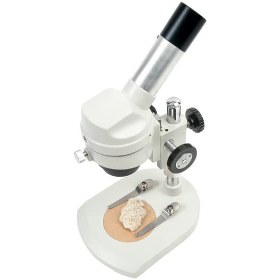
What is the low power objective lens on a microscope? Low power objectives cover a wide field of view and they are useful for examining large specimens or surveying many smaller specimens. This objective is useful for aligning the microscope. The power for the low objective is 10X. Place one of the prepared slides onto the stage of your microscope.
Is 40x a low power lens? Your microscope has 4 objective lenses: Scanning (4x), Low (10x), High (40x), and Oil Immersion (100x). … In addition to the objective lenses, the ocular lens (eyepiece) has a magnification.
What is the weakest objective lens? scanning lens. smallest and weakest objective lens (4x), but good for finding and focusing specimen. low power objective lens. small lens with low magnifying power (10x)
What is high power and low power on a microscope? When you change from low power to high power on a microscope, the high-power objective lens moves directly over the specimen, and the low-power objective lens rotates away from the specimen.
What is the low power objective lens on a microscope? – Related Questions
How do environmental scientists use microscopes?
Microscopes are used in environmental science to explore and investigate our planet and the life forms that inhabit (or inhabited) it. … Our upright microscopes are well suited for higher magnification observation of samples, especially microscopic organisms as in water treatment processes.
Which microscope uses the shortest wavelength?
The Scanning Electron Microscope (SEM) introduced here utilizes an electron beam whose wavelength is shorter than that of light and therefore observing a structure down to several nm in scale becomes possible.
What are slide holders in a microscope?
Containers such as slide boxes, trays, mailers, and carriers used to safely store and transport microscope slides; available in a variety of materials including cardboard, plastic, and stainless steel.
Are protists microscopic?
protist, any member of a group of diverse eukaryotic, predominantly unicellular microscopic organisms. They may share certain morphological and physiological characteristics with animals or plants or both.
How electrons move in an electron microscope?
The lenses are replaced by a series of coil-shaped electromagnets through which the electron beam travels. In an ordinary microscope, the glass lenses bend (or refract) the light beams passing through them to produce magnification. In an electron microscope, the coils bend the electron beams the same way.
How does the microscope works?
A simple light microscope manipulates how light enters the eye using a convex lens, where both sides of the lens are curved outwards. When light reflects off of an object being viewed under the microscope and passes through the lens, it bends towards the eye. This makes the object look bigger than it actually is.
What is the meaning of diaphragm on a microscope?
The microscope diaphragm, also known as the iris diaphragm, controls the amount and shape of the light that travels through the condenser lens and eventually passes through the specimen by expanding and contracting the diaphragm blades that resemble the iris of an eye.
How do things appear under a microscope?
A microscope is an instrument that can be used to observe small objects, even cells. The image of an object is magnified through at least one lens in the microscope. This lens bends light toward the eye and makes an object appear larger than it actually is.
What are some common magnification levels of a light microscope?
The eyepiece of most light microscopes comes with a few standard levels of magnification which range from 5x to 10x (probably the most common), and all the way up to 15x and 20x.
How stuff works light microscope?
Light from a mirror is reflected up through the specimen, or object to be viewed, into the powerful objective lens, which produces the first magnification. The image produced by the objective lens is then magnified again by the eyepiece lens, which acts as a simple magnifying glass.
What do microscopes do to an image?
A microscope is an instrument that can be used to observe small objects, even cells. The image of an object is magnified through at least one lens in the microscope. This lens bends light toward the eye and makes an object appear larger than it actually is.
Who uses light microscopes?
light microscopes are used to study living cells and for regular use when relatively low magnification and resolution is enough. electron microscopes provide higher magnifications and higher resolution images but cannot be used to view living cells.
What does it mean to resolution objects on microscope?
In microscopy, the term ‘resolution’ is used to describe the ability of a microscope to distinguish detail. In other words, this is the minimum distance at which two distinct points of a specimen can still be seen – either by the observer or the microscope camera – as separate entities.
What is the microscopic structure of the esophagus?
The microscopic structure of esophagus. The lining of the esophagus consists of more than one layer of cells, and the surface layer consists of flat or squamous cell. It’s a good example of stratified squamous epithelium (SS).
What careers use microscopes?
Some of the major jobs or careers that are known for their frequent use of the microscope are forensic scientists, jewelers, gemologists, botanists, and microbiologists. An example of a career emphasis that would predominantly use microscopes are researchers for science and public health.
Who made the compound light microscope?
The Dutch spectacle maker Hans Janssen and his son Zacharias are generally credited with creating these compound microscopes. The two of them built what was probably the first compound microscope in the last decade of the 16th century. It had a magnification that could be adjusted between 3 and 9x.
How does the light change on the compound microscope?
It is possible to control the amount of light that shines through the slide by changing the size of the opening present in the stage. It is also possible to change the illumination by moving the mirror in microscopes that use one or by changing the brightness of the light source in the Illuminator.
What is the difference between macroscopic and microscopic observation?
The macroscopic level includes anything seen with the naked eye and the microscopic level includes atoms and molecules, things not seen with the naked eye. Both levels describe matter.
Can you observe a piece of paper under a microscope?
Observation. When the paper sample is viewed under the microscope (at low magnification) the fibers will have a thread-like appearance and may look like cotton (when cotton is observed with the naked-eye). However, by switching to a higher magnification, the fibers should become more clear.
What microscope can be used to examine dna?
To view the DNA as well as a variety of other protein molecules, an electron microscope is used. Whereas the typical light microscope is only limited to a resolution of about 0.25um, the electron microscope is capable of resolutions of about 0.2 nanometers, which makes it possible to view smaller molecules.
How did robert hooke develop his compound microscope?
To combat dark specimen images, Hooke designed an ingenious method of concentrating light on his specimens, as shown in the illustration. He passed light generated from an oil lamp through a water-filled glass flask to diffuse the light and provide a more even and intense illumination for the samples.

