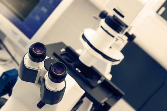
What is the low power objective on a compound microscope? Low power objectives cover a wide field of view and they are useful for examining large specimens or surveying many smaller specimens. This objective is useful for aligning the microscope. The power for the low objective is 10X.
What is the lowest objective on a compound microscope? The 4x objective lens has the lowest power and, therefore the highest field of view. As a result, it is easier to locate the specimen on the slide than if you start with a higher power objective.
What is the high power objective on a microscope? High Power Objective (40x): This objective (sometimes called the “high-dry” objective) is useful for observing fine detail such as the striations in skeletal muscle, the arrangement of Haversian systems in compact bone, types of nerve cells in the retina, etc.
What magnification is 40x objective? For example, a biological microscope with 10x eyepieces and a 40x objective has 400x magnification.
What is the low power objective on a compound microscope? – Related Questions
Do microscopic bugs live in your eyelashes?
Eyelash mites are microscopic organisms that naturally live at the base of your eyelashes. These tiny eight-legged eyelash bugs actually live in your hair follicles, and feed on dead skin cells and the oils in your skin. Eyelash mites are also known as demodex mites.
What is a monocular microscope?
Monocular microscopes, microscopes that are equipped with one eye piece, can magnify samples up to 1,000 times. If you need a microscope that magnifies at higher levels, a binocular microscope is right for you. Monocular microscopes are often used in classrooms and laboratories for observing slide samples.
How to measure magnification of microscope?
To calculate the total magnification of the compound light microscope multiply the magnification power of the ocular lens by the power of the objective lens. For instance, a 10x ocular and a 40x objective would have a 400x total magnification. The highest total magnification for a compound light microscope is 1000x.
What reflects light upwards on a microscope?
The simplest illuminator is a pivoted mirror to beam external light to the microscope. It’s used to direct room light, lamp light, or skylight from below the scope’s stage up through the specimen as transmitted light. … The flat side simply reflects light and gives a sharper image.
What is the drawback of use a light microscope?
d. Advantage: Light microscopes have high magnification. Electron microscopes are helpful in viewing surface details of a specimen. Disadvantage: Light microscopes can be used only in the presence of light and have lower resolution.
What is macroscopic and microscopic quantities?
While microscopic quantities represent a certain state in -space, their macroscopic counterparts are averages over -space. As a consequence, their dependency restricts to -space. Macroscopic quantities are obtained by the integration of the according microscopic quantity multiplied by the distribution function .
What is the function of scanning electron microscope?
A scanning electron microscope (SEM) scans a focused electron beam over a surface to create an image. The electrons in the beam interact with the sample, producing various signals that can be used to obtain information about the surface topography and composition.
How to properly use a dissecting microscope?
Lower the microscope body to its lowest point with the focusing knob on the sides of the microscope arm. Use the focus knob to raise the microscope body until the specimen image is the sharpest. b. Compensate for any differences in strength between your eyes.
Which scientist is credited to discover the microscope?
Zacharias Janssen, credited with inventing the microscope. (Image credit: Public domain.) For millennia, the smallest thing humans could see was about as wide as a human hair. When the microscope was invented around 1590, suddenly we saw a new world of living things in our water, in our food and under our nose.
Who first observed bacteria under the microscope?
Two men are credited today with the discovery of microorganisms using primitive microscopes: Robert Hooke who described the fruiting structures of molds in 1665 and Antoni van Leeuwenhoek who is credited with the discovery of bacteria in 1676.
What does the 10x objective do on the microscope?
100X (this means that the image being viewed will appear to be 100 times its actual size).
Is phase contrast microscope?
Phase contrast is a light microscopy technique used to enhance the contrast of images of transparent and colourless specimens. It enables visualisation of cells and cell components that would be difficult to see using an ordinary light microscope.
Why do light microscopes have a blue light filters?
A typical use would be a blue filter when using low levels of light in a brightfield application. (Like dry layer blood analysis.) When low levels of light are used, you usually get a yellowing effect because the lamp is turned low. The blue filter brings the light back to a more natural white light.
What does a staph infection look like under microscope?
The name Staphylococcus comes from the Greek staphyle, meaning a bunch of grapes, and kokkos, meaning berry, and that is what staph bacteria look like under the microscope, like a bunch of grapes or little round berries.
Why are cells typically microscopic in size?
The important point is that the surface area to the volume ratio gets smaller as the cell gets larger. Thus, if the cell grows beyond a certain limit, not enough material will be able to cross the membrane fast enough to accommodate the increased cellular volume. … That is why cells are so small.
What is the apparent size produced by a microscope called?
Optical magnification is the ratio between the apparent size of an object (or its size in an image) and its true size, and thus it is a dimensionless number. Optical magnification is sometimes referred to as “power” (for example “10× power”), although this can lead to confusion with optical power.
How many times magnification is an electron microscope have?
This makes electron microscopes more powerful than light microscopes. A light microscope can magnify things up to 2000x, but an electron microscope can magnify between 1 and 50 million times depending on which type you use!
Why are microscopes important to forensic scientists?
Microscopes are used throughout the modern forensic laboratory. They are essential in searching for evidence. They aid the examiner in identifying and comparing trace evidence. As the scales of justice symbolize forensic science, microscopes symbolize the trace evidence examiner.
What are the two main kinds of light microscopes?
used to view specimens are both simple and compound light microscopes, all using lenses. The difference is simple light microscopes use a single lens for magnification while compound lenses use two or more lenses for magnifications.
Who made the microscope better?
In 1676, Dutch cloth merchant-turned-scientist Antony van Leeuwenhoek further improved the microscope with the intent of looking at the cloth that he sold, but inadvertently made the groundbreaking discovery that bacteria exist.
Which muscle types appear striated under a microscope?
Skeletal muscles are long and cylindrical in appearance; when viewed under a microscope, skeletal muscle tissue has a striped or striated appearance.

