What is the microscopic anatomical makeup of visceral muscles? Each visceral muscle cell is long and thin with a single central nucleus and many protein fibers. The protein fibers are arranged into strings called intermediate filaments and masses known as dense bodies. Intermediate filaments contract to pull the dense bodies together and contract the visceral muscle cell.
What is the microscopic anatomical make up of? Microscopic anatomy includes cytology, the study of cells and histology, the study of tissues.
What is the microscopic anatomical makeup of skeletal muscles? The plasma membrane of the skeletal muscle fiber is called a sarcolemma. The muscle fiber contains long cylindrical structures, the myofibrils. The myofibrils almost entirely fill the cell and push the nuclei to the outer edges of the cell under the sarcolemma.
What contains visceral muscle? Visceral, or smooth, muscle is found inside organs such as the stomach and intestines, as well as in blood vessels.
What is the microscopic anatomical makeup of visceral muscles? – Related Questions
Can’t see anything in microscope?
If you cannot see anything, move the slide slightly while viewing and focusing. If nothing appears, reduce the light and repeat step 4. Once in focus on low power, center the object of interest by moving the slide. Rotate the objective to the medium power and adjust the fine focus only.
How to test sperm under a microscope?
To do a home test, a man would have to wait for around five minutes after ejaculation for the semen to liquefy, then apply a small amount to a plastic sheet and press it against the microscope for inspection. This can be done without getting semen on to the phone, says Kobori.
Who developed the first compound light microscope?
The Dutch spectacle maker Hans Janssen and his son Zacharias are generally credited with creating these compound microscopes. The two of them built what was probably the first compound microscope in the last decade of the 16th century.
What are the 4 different types of microscopes?
There are several different types of microscopes used in light microscopy, and the four most popular types are Compound, Stereo, Digital and the Pocket or handheld microscopes.
When was the electron microscope developed?
Ernst Ruska, a German electrical engineer, is credited with inventing the electron microscope. The earliest electron microscope was developed in 1931, and the first commercial, mass-produced instrument became available in 1939.
What power objective should your microscope be stored at?
Always place the 4X objective over the stage and be sure the stage is at its lowest position before putting the microscope away.
Do transmission electron microscopes use visible light?
Electron microscopes use a beam of electrons rather than visible light to illuminate the sample. They focus the electron beam using electromagnetic coils instead of glass lenses (as a light microscope does) because electrons can’t pass through glass.
Where are the slides placed on a microscope?
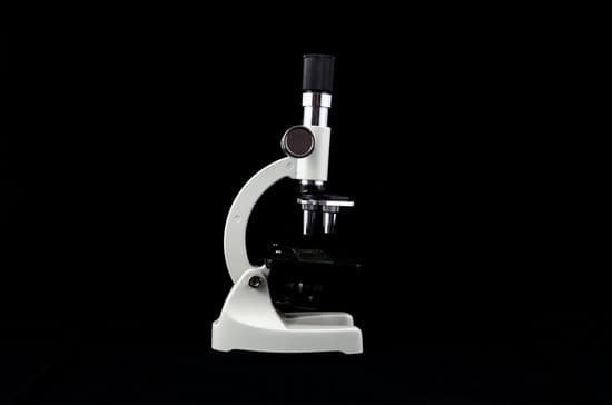
Stage: The flat platform where you place your slides. Stage clips hold the slides in place. Revolving Nosepiece or Turret: This is the part that holds two or more objective lenses and can be rotated to easily change power. Objective Lenses: Usually you will find 3 or 4 objective lenses on a microscope.
Who invented the inverted microscope?
An inverted microscope is a microscope with its light source and condenser on the top, above the stage pointing down, while the objectives and turret are below the stage pointing up. It was invented in 1850 by J. Lawrence Smith, a faculty member of Tulane University (then named the Medical College of Louisiana).
How is compound microscope used today?
Compound microscopes are used to view small samples that can not be identified with the naked eye. These samples are typically placed on a slide under the microscope. When using a stereo microscope, there is more room under the microscope for larger samples such as rocks or flowers and slides are not required.
How the microscope changed over time?
Microscopes became more stable and smaller. Lens improvements solved many of the optical problems that were common in earlier versions. The history of the microscope widens and expands from this point with people from around the world working on similar upgrades and lens technology at the same time.
How to adjust the interpupillary distance on a binocular microscope?
If you look through the eyepieces and see two images, the interpupillary distance is not correct. To correct it, slide the eyepieces closer together or farther apart until the two fields merge to form a single circle of light.
How does the compound microscope help us?
Compound microscopes can magnify specimens enough so that the user can see cells, bacteria, algae, and protozoa. You cannot see viruses, molecules, or atoms using a compound microscope because they are too small; an electron microscope is necessary to image such things.
Can atoms be observed under a microscope?
It’s tiny, but it’s visible. Atoms are so small that it’s almost impossible to see them without microscopes.
How did the microscope help scientists form the cell theory?
How did improvements in the microscope help scientists form the cell theory? Improvements allowed scientists to see cells in greater and greater detail and enabled them to discover cells in all types of living matter. … Eukaryotic cells have a nucleus and membrane-bound organelles, prokaryotic cells do not.
What kind of microscopes are used today?
There are several different types of microscopes used in light microscopy, and the four most popular types are Compound, Stereo, Digital and the Pocket or handheld microscopes.
What can you expect to see with a light microscope?
Explanation: You can see most bacteria and some organelles like mitochondria plus the human egg. You can not see the very smallest bacteria, viruses, macromolecules, ribosomes, proteins, and of course atoms.
Why do electron microscopes have higher resolving power?
Electron microscopes differ from light microscopes in that they produce an image of a specimen by using a beam of electrons rather than a beam of light. Electrons have much a shorter wavelength than visible light, and this allows electron microscopes to produce higher-resolution images than standard light microscopes.
How to calculate the magnification of a compound microscope?
To calculate the total magnification of the compound light microscope multiply the magnification power of the ocular lens by the power of the objective lens. For instance, a 10x ocular and a 40x objective would have a 400x total magnification. The highest total magnification for a compound light microscope is 1000x.
Do objects viewed under the microscope appear upside down?
A microscope is an instrument that magnifies an object. … A specimen that is right-side up and facing right on the microscope slide will appear upside-down and facing left when viewed through a microscope, and vice versa.
Where does the history of microscope begun?
Spectacles first made in Italy. Two Dutch spectacle-makers and father-and-son team, Hans and Zacharias Janssen, create the first microscope. Robert Hooke’s famous “Micrographia” is published, which outlines Hooke’s various studies using the microscope.

