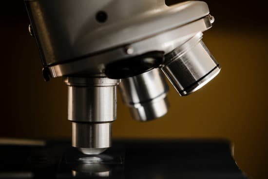
What is the proper way to transport a microscope? When transporting the microscope, hold it in an upright position with one hand on its arm and the other supporting its base. Avoid jarring the instrument when setting it down. Use only special grit-free lens paper to clean the lenses.
How do you transport a microscope? WHEN TRANSPORTING THE MICROSCOPE, HOLD IT IN AN UPRIGHT POSITION WITH ONE HAND ON ITS ARM AND THE OTHER SUPPORTING ITS BASE. AVOID SWINGING THE INSTRUMENT DURING ITS TRANSPORT AND JARRING THE INSTRUMENT WHEN SETTING IT DOWN. THE MICROSCOPE LENS MAY BE CLEANED (WITH ANY SOFT TISSUE).
What will you do to transport the microscope safely? Place one hand on the bottom of the microscope and use the back arm of the microscope for carrying it. The image below shows the correct hand holds for safely moving a microscope without damaging it. Always use two hands when moving the microscope. Ensure the microscope is not plugged in before moving it.
How should a microscope be carried safely and stored correctly? When carrying the microscope, always use two hands with one hand supporting the base and theother hand holding the arm. Properly store the microscope by lowering the nosepiece, turning off the light source, and placing the objective lenses on the lowest setting. Cover with a dust jacket.
What is the proper way to transport a microscope? – Related Questions
Who started microscope?
The development of the microscope allowed scientists to make new insights into the body and disease. It’s not clear who invented the first microscope, but the Dutch spectacle maker Zacharias Janssen (b. 1585) is credited with making one of the earliest compound microscopes (ones that used two lenses) around 1600.
What is the substage condenser on a microscope?
The substage condenser gathers light from the microscope light source and concentrates it into a cone of light that illuminates the specimen with uniform intensity over the entire viewfield. … The size and numerical aperture of the light cone is determined by adjustment of the aperture diaphragm.
How to find dust mites microscope?
Dust mites are too small to see with the human eye, but can be seen at 20 times magnification with a microscope. You can easily calculate the total magnification of your microscope to see if it is strong enough to see the mite by multiplying the eyepiece magnification by the objective lens modification.
Why is it important the microscope be covered?
When the microscope is not in use keep it covered with the dust cover. This alone will extend the life of your microscope. … This can allow dust to collect within the eye tubes, which can be difficult to clean.
What is the difference between microscopic and macroscopic current?
Microscopic approach considers the behaviour of every molecule by using statistical methods. In Macroscopic approach we are concerned with the gross or average effects of many molecules’ infractions. These effects, such as pressure and temperature, can be perceived by our senses and can be measured with instruments.
When would you use a confocal microscope?
As a distinctive feature, confocal microscopy enables the creation of sharp images of the exact plane of focus, without any disturbing fluorescent light from the background or other regions of the specimen. Therefore, structures within thicker objects can be conveniently visualized using confocal microscopy.
Which has a greater resolution compound light or electron microscope?
Electron microscopes differ from light microscopes in that they produce an image of a specimen by using a beam of electrons rather than a beam of light. Electrons have much a shorter wavelength than visible light, and this allows electron microscopes to produce higher-resolution images than standard light microscopes.
Did robert hooke invented the first microscope?
Although Hooke did not make his own microscopes, he was heavily involved with the overall design and optical characteristics. The microscopes were actually made by London instrument maker Christopher Cock, who enjoyed a great deal of success due to the popularity of this microscope design and Hooke’s book.
What year was the microscope invented?
Lens Crafters Circa 1590: Invention of the Microscope. Every major field of science has benefited from the use of some form of microscope, an invention that dates back to the late 16th century and a modest Dutch eyeglass maker named Zacharias Janssen.
Do high schools use microscopes?
These are the most popular student microscopes used in high schools around the world. High school compound microscopes will always have magnification of 40x, 100x, and 400x. Many of the high school microscopes will also have 1000x magnification.
What is microscopic examination of metals?
During Microstructure Analysis of metals and alloys, a Microscopic Examination is conducted to study the microstructural features of the material under magnification.
How to focus an electron microscope?
A positively charged electrode (anode) attracts the electrons and accelerates them into an energetic beam. An electromagnetic coil brings the electron beam to a very precise focus, much like a lens. Another coil, lower down, steers the electron beam from side to side.
What focus on a microscope is used at high magnification?
Use ONLY the fine focus control when focusing the higher power objectives (20X, 40X, 100X) on a slide. The course focus control is too course for focusing with these objectives.
What was the first microscope like?
The early simple “microscopes” which were really only magnifying glasses had one power, usually about 6X – 10X . One thing that was very common and interesting to look at was fleas and other tiny insects. These early magnifiers were hence called “flea glasses”.
How lenses of microscope works?
A simple light microscope manipulates how light enters the eye using a convex lens, where both sides of the lens are curved outwards. When light reflects off of an object being viewed under the microscope and passes through the lens, it bends towards the eye. This makes the object look bigger than it actually is.
Is the ocular lens in light microscopes?
Ordinary light microscopes are equipped with three objective lenses (5 ×/10 ×, 40 ×, and 90/100 ×), and two ocular (5 ×, 10 ×) lenses.
What is the magnification of microscope objectives?
Objectives typically have magnifying powers that range from 1:1 (1X) to 100:1 (100X), with the most common powers being 4X (or 5X), 10X, 20X, 40X (or 50X), and 100X.
What type of microscope to view flagella?
The flagella stain allows observation of bacterial flagella under the light microscope. Bacterial flagella are normally too thin to be seen under such conditions. The flagella stains employs a mordant to coat the flagella with stain until they are thick enough to be seen.
What does a microscope used for?
A microscope is an instrument that can be used to observe small objects, even cells. The image of an object is magnified through at least one lens in the microscope. This lens bends light toward the eye and makes an object appear larger than it actually is.
Can chromosome be seen under a microscope?
Chromosomes are not visible in the cell’s nucleus—not even under a microscope—when the cell is not dividing. However, the DNA that makes up chromosomes becomes more tightly packed during cell division and is then visible under a microscope.
What does a pointer do in a microscope?
Pointer: A piece of high tensile wire that sits in the eyepiece and enables a viewer to point at a specific area of a specimen.

