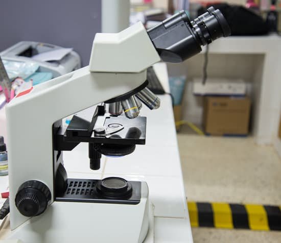
What is the purpose of the light microscope? Principles. The light microscope is an instrument for visualizing fine detail of an object. It does this by creating a magnified image through the use of a series of glass lenses, which first focus a beam of light onto or through an object, and convex objective lenses to enlarge the image formed.
Why do we need light microscopes to look at cells? Because most cells are too small to be seen by the naked eye, the study of cells has depended heavily on the use of microscopes. … Thus, the cell achieved its current recognition as the fundamental unit of all living organisms because of observations made with the light microscope.
Why do scientist use microscopes to study cells? A cell is the smallest unit of life. Most cells are so tiny that they cannot be seen with the naked eye. Therefore, scientists use microscopes to study cells. Electron microscopes provide higher magnification, higher resolution, and more detail than light microscopes.
What is the purpose of using a light microscope? The light microscope is an instrument for visualizing fine detail of an object. It does this by creating a magnified image through the use of a series of glass lenses, which first focus a beam of light onto or through an object, and convex objective lenses to enlarge the image formed.
What is the purpose of the light microscope? – Related Questions
What organelles can be seen with a transmission electron microscope?
The cell wall, nucleus, vacuoles, mitochondria, endoplasmic reticulum, Golgi apparatus, and ribosomes are easily visible in this transmission electron micrograph.
How does light move through a compound light microscope?
Microscopes are effectively just tubes packed with lenses, curved pieces of glass that bend (or refract) light rays passing through them. … When light shines on the specimen at the bottom, it travels straight through or reflects off the surface, passing up through the lenses into the eyepiece.
Can you see molecules with a microscope?
This, believe it or not, is a microscope. It can help us see very small particles like molecules by feeling the particle with the tip of its needle. These very powerful microscopes are called atomic force microscopes, because they can see things by feeling the forces between atoms. …
Do cardiac muscles appear striated under a microscope?
Cardiac muscle tissue, like skeletal muscle tissue, looks striated or striped. The bundles are branched, like a tree, but connected at both ends.
What is the difference between a hand lens and microscope?
The difference between hand lens and microscope magnifications comes from the number of lenses. With a magnifying glass or hand lens, the magnification is limited to the single lens. Since the lens has one focal length from the lens to the focus point, the magnification is fixed.
What is the function of the handle on a microscope?
Part of the handle used to lift and carry it. Base The bottom of the scope. Usually houses the light source, if one is present. The extended, rear portion of the base also functions as the handle and is used to lift and carry the microscope.
What advantage is there to using a parfocal microscope?
A parfocal zoom lens maintains focus as the focal point changes and the lens is zoomed (changing both focal length and magnification). A parfocal lens allows for more accurate focusing at the maximum focal length, and then quick zooming back to a shorter focal length.
What is the smallest thing seen with a microscope?
The smallest thing that we can see with a ‘light’ microscope is about 500 nanometers. A nanometer is one-billionth (that’s 1,000,000,000th) of a meter. So the smallest thing that you can see with a light microscope is about 200 times smaller than the width of a hair. Bacteria are about 1000 nanometers in size.
How does phase microscope work?
Phase contrast microscopy translates small changes in the phase into changes in amplitude (brightness), which are then seen as differences in image contrast. Unstained specimens that do not absorb light are known as phase objects. … This allows the specimen to be illuminated by parallel light that has been defocused.
Who invented travelling microscope?
It’s called a Withering microscope, after a medical student called William Withering who designed a simple pocket microscope, made of brass, to help him and others in the study of botany when out of the laboratory.
How does the light microscope magnify image simple?
Principles. The light microscope is an instrument for visualizing fine detail of an object. It does this by creating a magnified image through the use of a series of glass lenses, which first focus a beam of light onto or through an object, and convex objective lenses to enlarge the image formed.
What is the resolving power of a typical electron microscope?
The resolution limit of light microscopes is about 200nm, the maximum useful magnification a light microscope can provide is about 1,000x. The resolution limit of electron microscopes is about 0.2nm, the maximum useful magnification an electron microscope can provide is about 1,000,000x.
What is the function of a condenser microscope?
On upright microscopes, the condenser is located beneath the stage and serves to gather wavefronts from the microscope light source and concentrate them into a cone of light that illuminates the specimen with uniform intensity over the entire viewfield.
What is the difference between microscopic and macroscopic quizlet?
What is the difference between macroscopic and microscopic views? Macroscopic can be observed with the eye, whereas microscopic is seen through the use of a microscope and can not be seen with the naked eye.
What can we see with a light microscope?
Explanation: You can see most bacteria and some organelles like mitochondria plus the human egg. You can not see the very smallest bacteria, viruses, macromolecules, ribosomes, proteins, and of course atoms.
What focus should we start with the microscopes?
When focusing on a slide, ALWAYS start with either the 4X or 10X objective. Once you have the object in focus, then switch to the next higher power objective. Re-focus on the image and then switch to the next highest power.
How has the discovery of the microscope helped society?
A microscope lets the user see the tiniest parts of our world: microbes, small structures within larger objects and even the molecules that are the building blocks of all matter. The ability to see otherwise invisible things enriches our lives on many levels.
How to focus a microscope fine and coarse adjustment?
Slowly raise the stage until you are roughly in focus. Rotate the turret Clockwise to Higher Powered Objectives and re-focus using the Fine Focus Knob. For each rotation, you’ll want to re-adjust focus using the fine focus knob only. This is to ensure the objective doesn’t bump up against the slide too harshly.
Can microscopic colitis cause hair loss?
Anecdotally hair loss is commonly reported by patients with IBD; however the exact cause, prevalence, and relationship to IBD medications and disease activity are poorly defined. Previously, a retrospective case series in patients with ulcerative colitis (UC) described an overall low prevalence of hair loss.
How do we calculate the total magnification of a microscope?
The total magnification of the microscope is calculated from the magnifying power of the objective multiplied by the magnification of the eyepiece and, where applicable, multiplied by intermediate magnifications. A distinction is made between magnification and lateral magnification.

