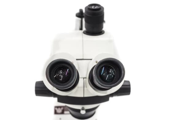
What is the resolution for s electron microscope? The wavelength of electrons is much smaller than that of photons (2.5 pm at 200 keV). Thus the resolution of an electron microscope is theoretically unlimited for imaging cellular structure or proteins. Practically, the resolution is limited to ~0.1 nm due to the objective lens system in electron microscopes.
What is the best resolution for an electron microscope? A scanning transmission electron microscope has achieved better than 50 pm resolution in annular dark-field imaging mode and magnifications of up to about 10,000,000× whereas most light microscopes are limited by diffraction to about 200 nm resolution and useful magnifications below 2000×.
What is the resolution of an electron microscope in micrometers? Center for Materials Research and Analysis. University of Nebraska at Lincoln. “Electron Microscopes were developed due to the limitations of Light Microscopes which are limited by the physics of light to 500x or 1000x magnification and a resolution of 0.2 micrometers.”
Do electron microscopes have high resolution? At present, the highest point resolution realised in phase contrast transmission electron microscopy is around 0.5 ångströms (0.050 nm).
What is the resolution for s electron microscope? – Related Questions
What is the microscopic study of cells?
“cell”) are professionals who study cells via microscopic examinations and other laboratory tests. They are trained to determine which cellular changes are within normal limits and which are abnormal.
What is meant by dissecting microscope?
A dissecting microscope is used to view three-dimensional objects and larger specimens, with a maximum magnification of 100x. This type of microscope might be used to study external features on an object or to examine structures not easily mounted onto flat slides.
Can atoms be seen by a microscope?
Atoms are really small. So small, in fact, that it’s impossible to see one with the naked eye, even with the most powerful of microscopes. … Now, a photograph shows a single atom floating in an electric field, and it’s large enough to see without any kind of microscope.
Can atoms be seen with microscope?
Atoms are really small. So small, in fact, that it’s impossible to see one with the naked eye, even with the most powerful of microscopes. … Now, a photograph shows a single atom floating in an electric field, and it’s large enough to see without any kind of microscope.
What is the diaphragm used for in a light microscope?
Iris Diaphragm controls the amount of light reaching the specimen. It is located above the condenser and below the stage. Most high quality microscopes include an Abbe condenser with an iris diaphragm. Combined, they control both the focus and quantity of light applied to the specimen.
Can interstitial cystitis cause microscopic hematuria?
Interstitial cystitis (IC) is an ill-defined, chronic inflammatory condition of the bladder of unknown etiology characterized by pelvic pain, frequency, urgency, and nocturia. These patients classically present with a constellation of urologic complaints, which may include microscopic and gross hematuria.
What is the main function of the microscope?
A microscope is an instrument that can be used to observe small objects, even cells. The image of an object is magnified through at least one lens in the microscope. This lens bends light toward the eye and makes an object appear larger than it actually is.
What is wbc in microscopic examination?
The number of WBCs in urine sediment is normally low. When the number is high, it indicates an infection or inflammation somewhere in the urinary tract. Women especially must take care during specimen collection so that vaginal secretions (that can be high in WBCs) don’t contaminate the urine.
Does the electron microscope allow scientists to observe viruses?
12/05/20 The electron microscope allows scientists to look at even the smallest structures. For example, it provides detailed images of viruses and crystal lattices. There is always high vacuum inside the device.
How to remove microscope with hardened oil?
If you need to remove immersion oil that has been left on a lens and hardened, moisten lens paper with a small amount of xylene or microscope lens cleaning solution. Gently wipe lens surfaces, allowing enough time for the solution to soften the hardened oil. Once oil is removed wipe surfaces again.
When was a microscope made?
In around 1590, Hans and Zacharias Janssen had created a microscope based on lenses in a tube [1]. No observations from these microscopes were published and it was not until Robert Hooke and Antonj van Leeuwenhoek that the microscope, as a scientific instrument, was born.
How much is a transmission electron microscope cost?
The cost of a transmission electron microscope (TEM) can range from $300,000 to $10,000,000. The cost of a focused ion beam electron microscope (FIB) can range from $500,000 to $4,000,000. There can be a high degree of variation in the cost of an electron microscope between manufacturers and models.
How was invented the microscope?
A Dutch father-son team named Hans and Zacharias Janssen invented the first so-called compound microscope in the late 16th century when they discovered that, if they put a lens at the top and bottom of a tube and looked through it, objects on the other end became magnified.
How is the total magnification of a microscope image calculated?
The first is the objective lens, a highly magnified lens with between 40 and 100 times magnification that produces a high-resolution re-creation of the original image. … In other words, the total magnification of a microscope is understood as the magnification of the objective lens multiplied by that of the optical lens.
What does a scabies mite look like under a microscope?
What scabies look like. Scabies is caused by the mite known as the Sarcoptes scabiei. These mites are so tiny that they can’t be seen by the human eye. When viewed by a microscope, you’d see they have a round body and eight legs.
When was the microscope?
Zacharias Janssen, credited with inventing the microscope. (Image credit: Public domain.) For millennia, the smallest thing humans could see was about as wide as a human hair. When the microscope was invented around 1590, suddenly we saw a new world of living things in our water, in our food and under our nose.
What is high resolution microscope?
High-resolution transmission electron microscopy is an imaging mode of specialized transmission electron microscopes that allows for direct imaging of the atomic structure of samples. … At present, the highest point resolution realised in phase contrast transmission electron microscopy is around 0.5 ångströms (0.050 nm).
What is microscope slide meaning?
microscope slide Add to list Share. Definitions of microscope slide. a small flat rectangular piece of glass on which specimens can be mounted for microscopic study.
What parts are located in the body of the microscope?
Body tube (Head): The body tube connects the eyepiece to the objective lenses. Arm: The arm connects the body tube to the base of the microscope. Coarse adjustment: Brings the specimen into general focus. Fine adjustment: Fine tunes the focus and increases the detail of the specimen.
What is the microscopic air sacs in the lungs called?
Listen to pronunciation. (al-VEE-oh-ly) Tiny air sacs at the end of the bronchioles (tiny branches of air tubes in the lungs). The alveoli are where the lungs and the blood exchange oxygen and carbon dioxide during the process of breathing in and breathing out.
What type of microscope is best for viewing yeast cells?
For one thing, yeast and buds can be seen under a high magnification (1000x) bright field microscope, such as a compound microscope. This allows us to see oval shaped microscopic bodies, which are the yeast cells’ units of protoplasm.

