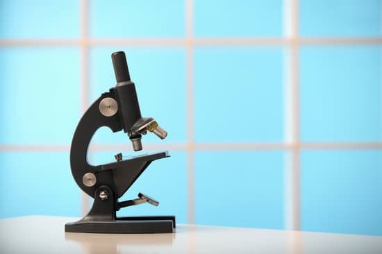
What is the revolving nosepiece used for on a microscope? Revolving Nosepiece or Turret: This is the part that holds two or more objective lenses and can be rotated to easily change power. Objective Lenses: Usually you will find 3 or 4 objective lenses on a microscope. They almost always consist of 4X, 10X, 40X and 100X powers.
What is a revolving nosepiece? The revolving nosepiece is the inclined, circular metal plate to which the objective lenses, usually four, are attached. … The final magnification is the product of the magnification of the ocular and objective lenses. A slide is placed on the mechanical stage and is moved by rotating the stage control knobs.
What is the purpose of nosepiece? Nosepiece: This circular structure is where the different objective lenses are screwed in. To change the magnification power, simply rotate the turret. Objective Lenses: Usually you will find 3 or 4 objective lenses on a microscope.
Where is the rotating nose piece on a microscope? The revolving nosepiece is located at bottom of the head of a microscope on an upright microscope.
What is the revolving nosepiece used for on a microscope? – Related Questions
Is microscopic blood common with uti?
Symptoms can include a persistent urge to urinate, pain and burning with urination, and extremely strong-smelling urine. For some people, especially older adults, the only sign of illness might be microscopic blood in the urine. Kidney infections (pyelonephritis).
What part of the microscope focuses light onto the specimen?
Condenser Lens – This lens system is located immediately under the stage and focuses the light on the specimen.
How is the total magnification of the compound microscope determined?
The total magnification is calculated by multiplying the magnification of the ocular lens by the magnification of the objective lens. … Compound microscopes usually include exchangeable objective lenses with different magnifications (e.g 4x, 10x, 40x and 60x), mounted on a turret, to adjust the magnification.
What are the two types of electron microscopes?
Today there are two major types of electron microscopes used in clinical and biomedical research settings: the transmission electron microscope (TEM) and the scanning electron microscope (SEM); sometimes the TEM and SEM are combined in one instrument, the scanning transmission electron microscope (STEM):
Why are light microscopes better than electron?
Resolution: The biggest advantage is that they have a higher resolution and are therefore also able of a higher magnification (up to 2 million times). Light microscopes can show a useful magnification only up to 1000-2000 times. This is a physical limit imposed by the wavelength of the light.
Why would a wet mount shake or vibrate under microscope?
Camera vibrations can become a problem if the exposure time is too long. … Small specimens which are surrounded by water water are able to float around between the slide and cover glass and will start to vibrate when one is tapping on the table on which the microscope stands.
What can i eat if i have microscopic colitis?
Avoid beverages that are high in sugar or sorbitol or contain alcohol or caffeine, such as coffee, tea and colas, which may aggravate your symptoms. Choose soft, easy-to-digest foods. These include applesauce, bananas, melons and rice. Avoid high-fiber foods such as beans and nuts, and eat only well-cooked vegetables.
How to focus a compound light microscope?
Look at the objective lens (3) and the stage from the side and turn the focus knob (4) so the stage moves upward. Move it up as far as it will go without letting the objective touch the coverslip. Look through the eyepiece (1) and move the focus knob until the image comes into focus.
What is the microscopic structure of bone and cartilage?
Haversian canals contain blood vessels, and are interconnected with the marrow cavity by transverse, Volkmann’s canals. The interstitial substance contains lacunae, which contain os-teocytes. Spongy bone is also composed of lamellae with lacunae in the interstitial substance.
What type of microscope do we use in biology?
Compound light microscopes are one of the most familiar of the different types of microscopes as they are most often found in science and biology classrooms.
How many objectives they are on the virtual microscope?
The microscope turret can simulate five different objectives, with the current objective highlighted. Click on a new objective to change the current magnification.
Why do microscopes invert images?
Under the slide on which the object is being magnified, there is a light source that shines up and helps you to see the object better. This light is then refracted, or bent around the lens. Once it comes out of the other side, the two rays converge to make an enlarged and inverted image.
How to determine the magnifying power of a microscope?
To calculate the total magnification of the compound light microscope multiply the magnification power of the ocular lens by the power of the objective lens. For instance, a 10x ocular and a 40x objective would have a 400x total magnification. The highest total magnification for a compound light microscope is 1000x.
Does microscopic colitis cause fatigue?
Other symptoms of microscopic colitis could include fever, joint pain, and fatigue. 4 These symptoms may be a result of the inflammatory process that is part of an autoimmune or immune-mediated disease.
What is the structure of a dissecting microscope?
It produces a three-dimensional image of the specimen rather than a flat image. Dissecting microscope contains two separate objective lens and eyepiece, which creates two separate optical paths for each eye. As a result, it creates a 3D image of the specimen.
Do we still use microscopes to look at cells?
The light microscope remains a basic tool of cell biologists, with technical improvements allowing the visualization of ever-increasing details of cell structure. Contemporary light microscopes are able to magnify objects up to about a thousand times.
How can the resolution of the microscope be improved?
The resolution of a specimen viewed through a microscope can be increased by changing the objective lens. The objective lenses are the lenses that protrude downward over the specimen. … Rotate the nose piece so that the shortest objective lens is positioned over the slide.
Who created the prototype of the modern scanning electron microscope?
However, the principles of the electron microscope are still based on the first prototype that was developed by Ernst Ruska. Electron microscopes have surpassed many of the limitations of optical microscopes, with improved resolution that makes it possible to view microscopic objects such as atoms.
Are microscopes bad for eyes?
The narrow field of view from most microscope eyepieces is a major cause of eye strain and bad posture. Users who wear spectacles often have to remove them, increasing the risk of eye strain; and many users also suffer the distraction of floating fragments of tissue debris in the eye.
What limits resolution of optical microscope?
The resolution of the light microscope cannot be small than the half of the wavelength of the visible light, which is 0.4-0.7 µm. When we can see green light (0.5 µm), the objects which are, at most, about 0.2 µm.
How to adjust microscope?
Use the coarse and fine focus control knobs to adjust the focus of the sample. When the sample is clearly visible, use only your left eye. Do NOT adjust the focus knobs. Instead, adjust the diopter on the left eyepiece until the sample comes clearly into view.

