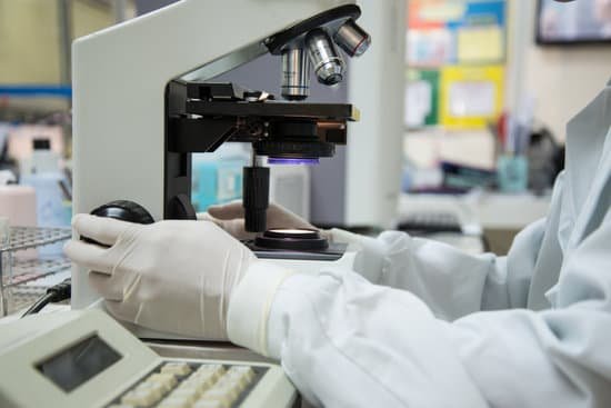
What is the smallest object visible with a light microscope? The smallest thing that we can see with a ‘light’ microscope is about 500 nanometers. A nanometer is one-billionth (that’s 1,000,000,000th) of a meter. So the smallest thing that you can see with a light microscope is about 200 times smaller than the width of a hair. Bacteria are about 1000 nanometers in size.
What microscope is used to see very small objects? Viewable objects by magnification.
What are smallest cell structures that can be seen by the light microscope? In practical terms, bacteria and mitochondria, which are about 500 nm (0.5 μm) wide, are generally the smallest objects whose shape can be clearly discerned in the light microscope; details smaller than this are obscured by effects resulting from the wave nature of light.
What is the smallest thing ever seen? Scientists have taken the first ever snapshot of an atom’s shadow—the smallest ever photographed using visible light.
What is the smallest object visible with a light microscope? – Related Questions
Will put under the microscope?
put (someone or something) under a microscope. To begin closely inspecting someone or something; to examine someone or something with intense scrutiny.
How to measure field diameter of microscope?
For instance, if your eyepiece reads 10X/22, and the magnification of your objective lens is 40. First, multiply 10 and 40 to get 400. Then divide 22 by 400 to get a FOV diameter of 0.055 millimeters.
How many lenses does a scanning electron microscope have?
The electron beam, which typically has an energy ranging from 0.2 keV to 40 keV, is focused by one or two condenser lenses to a spot about 0.4 nm to 5 nm in diameter.
What is meant by the working distance of a microscope?
■ Working Distance (W.D.) The distance between the front end of a microscope objective and the. surface of the workpiece at which the sharpest focusing is obtained.
When was the modern light microscope invented?
In around 1590, Hans and Zacharias Janssen had created a microscope based on lenses in a tube [1]. No observations from these microscopes were published and it was not until Robert Hooke and Antonj van Leeuwenhoek that the microscope, as a scientific instrument, was born.
Can images be inverted while using a dissecting microscope?
Binocular and dissecting microscopes will also not show an inverted image because of their increased level of magnification. … Even with an inverted image, microscopes can increase the magnification of an image phenomenally.
What is the definition for the word microscope?
1 : an optical instrument consisting of a lens or combination of lenses for making enlarged images of minute objects especially : compound microscope. 2 : a non-optical instrument (such as one using radiations other than light or using vibrations) for making enlarged images of minute objects an acoustic microscope.
What is typical magnification dissecting microscope?
Stereo microscopes are also called stereoscopic and dissecting microscopes. Compared to compound microscopes, dissecting microscopes have lower power optical systems and illuminators; a dissecting microscope’s typical magnification ranges from about 10x-40x.
What is the relationship between microscopic animals and plants?
Herbivory is an interaction in which a plant or portions of the plant are consumed by an animal. At the microscopic scale, herbivory includes the bacteria and fungi that cause disease as they feed on plant tissue. Microbes that break down dead plant tissue are also specialized herbivores.
What does zoom magnification microscope?
The term “zoom” refers to continuous viewing of magnification among a range, for example if the zoom range is 7x -45x, you would be able to view 7x, 8x, 9x, etc. all the way up to 45x. Stereo zoom microscopes have eyepieces for viewing samples, and some have a trinocular (camera) port as well.
What does an eyepiece lens do on a microscope?
The eyepiece, or ocular lens, is the part of the microscope that magnifies the image produced by the microscope’s objective so that it can be seen by the human eye.
Which lens is used in microscope and telescope?
The simplest compound microscope is constructed from two convex lenses (Figure 2.9. 1). The objective lens is a convex lens of short focal length (i.e., high power) with typical magnification from 5× to 100×. The eyepiece, also referred to as the ocular, is a convex lens of longer focal length.
What is the shortest objective called on a microscope?
After the light has passed through the specimen, it enters the objective lens (often called “objective” for short). The shortest of the three objectives is the scanning-power objective lens (N), and has a power of 4X.
What is confocal microscope resolution?
In practice, the best horizontal resolution of a confocal microscope is about 0.2 microns, and the best vertical resolution is about 0.5 microns.
How to measure in micrometers on a microscope?
Usually the stage micrometer is 1 millimeter in length and each stage micrometer division is equal to 0.01. So, if there are 80 divisions across your field of view then the diameter for that magnification level is 0.8 millimeters or 800 micrometers.
What part of the microscope regulates the amount of light?
Iris diaphragm dial: Dial attached to the condenser that regulates the amount of light passing through the condenser. The iris diaphragm permits the best possible contrast when viewing the specimen.
What microscope is used to see blood cells?
The compound microscope can be used to view a variety of samples, some of which include: blood cells, cheek cells, parasites, bacteria, algae, tissue, and thin sections of organs. Compound microscopes are used to view samples that can not be seen with the naked eye.
What is the specimens for a electron microscope?
Electron microscopes are used to investigate the ultrastructure of a wide range of biological and inorganic specimens including microorganisms, cells, large molecules, biopsy samples, metals, and crystals. Industrially, electron microscopes are often used for quality control and failure analysis.
Why is the electron microscope useful?
Electron microscopy (EM) is a technique for obtaining high resolution images of biological and non-biological specimens. It is used in biomedical research to investigate the detailed structure of tissues, cells, organelles and macromolecular complexes.
How to light microscopes differ from electron microscopes?
Electron microscopes differ from light microscopes in that they produce an image of a specimen by using a beam of electrons rather than a beam of light. Electrons have much a shorter wavelength than visible light, and this allows electron microscopes to produce higher-resolution images than standard light microscopes.
Which organisms can you see with a typical light microscope?
Explanation: You can see most bacteria and some organelles like mitochondria plus the human egg. You can not see the very smallest bacteria, viruses, macromolecules, ribosomes, proteins, and of course atoms.

