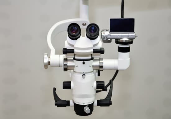
What kind of lens is used in camera and microscope? It is made of two convex lenses: the first, the ocular lens, is close to the eye; the second is the objective lens. Compound microscopes are much larger, heavier and more expensive than simple microscopes because of the multiple lenses.
What type of lens is used in camera? In a photographic camera, a convex lens of larger focal length is used.
What type of lens is used in microscope? Microscopes use convex lenses in order to focus light.
What types of lens is used in microscope and telescope? Convex or Converging lenses are used in microscopes and telescopes.
What kind of lens is used in camera and microscope? – Related Questions
Can chlamydia be seen under a microscope?
The discharge is usually clear and stringy. In a sexual health clinic, the doctor or nurse may take a specimen and look at this under the microscope. They are looking for signs of infection such as an increased amount of white blood cells, and the chlamydia bacteria.
What does urinalysis macroscopic reflex microscopic test for?
This test looks at a sample of your urine under a microscope. It can see cells from your urinary tract, blood cells, crystals, bacteria, parasites, and cells from tumors. This test is often used to confirm the findings of other tests or add information to a diagnosis.
Which organelles are visible with the light microscope quizlet?
How do we, scientists, know about these organelles? We observed different organelles under the compound light microscope; cytoplasm, cell wall, cell membrane, nucleus, chloroplast and something that could have been vacuoles or lysosomes.
Can i buy portable microscope?
American Optics, Sper Scientific, Electron Microscopy Sciences, and ElectroOptics all offer pen-sized microscopes for both student and professional use. Home Training Tools, Scientific Online, PEAK Optics and Carson all offer handheld and stand-alone portable microscopes with 25x to 100x magnification.
What types of scientist use microscopes?
Today, microscopes are used by all types of scientists, including cell biologists, microbiologists, virologists, forensic scientists, entomologists, taxonomists, and many other types.
Can you see dust mites without a microscope?
Are dust mites visible? The answer to the first question is quite simple. No, you cannot easily see dust mites. The reason being is because these organisms are so microscopic that they can’t be seen by the naked eye.
What power microscope do i need to see bacteria?
In order to actually see bacteria swimming, you’ll need a lens with at least a 400x magnification. A 1000x magnification can show bacteria in stunning detail. However, at a higher magnification, it can be increasingly difficult to keep them in focus as they move.
Can i cure my microscopic colitis?
Microscopic colitis may get better on its own. But when symptoms persist or are severe, you may need treatment to relieve them. Doctors usually try a stepwise approach, starting with the simplest, most easily tolerated treatments.
What is the function of an electron microscope?
The electron microscope uses a beam of electrons and their wave-like characteristics to magnify an object’s image, unlike the optical microscope that uses visible light to magnify images.
How do i find the total magnification on a microscope?
To figure the total magnification of an image that you are viewing through the microscope is really quite simple. To get the total magnification take the power of the objective (4X, 10X, 40x) and multiply by the power of the eyepiece, usually 10X.
What is a dissecting microscope best used for?
A dissecting microscope is used to view three-dimensional objects and larger specimens, with a maximum magnification of 100x. This type of microscope might be used to study external features on an object or to examine structures not easily mounted onto flat slides.
How to count microscope for hemocytometer?
To count cells using a hemocytometer, add 15-20μl of cell suspension between the hemocytometer and cover glass using a P-20 Pipetman. The goal is to have roughly 100-200 cells/square. Count the number of cells in all four outer squares divide by four (the mean number of cells/square).
What is the lowest magnification of a dissecting microscope?
Dissecting microscopes provide low magnification (usually 10x – 40x is a common range) and they are used to view detail in objects you can already see with the naked eye.
Can sperm cells be seen under a microscope?
The air-fixed, stained spermatozoa are observed under a bright-light microscope at 400x or 1000x magnification. Their viability and mor- phology can be analysed at the same time.
How to increase contrast on microscope?
Contrast may be improved by placing suitable apertures or filters within the optical path, either in the illuminating system alone (dark ground or Rheinberg illumination), or in conjugate planes in the imaging system (e.g. for phase contrast, differential interference contrast or polarised light microscopy).
How much does a raman microscope cost?
How much does a Raman microscope cost? Optosky Raman spectrometer prices range from USD39,800 to USD99,980 above.
Do modern microscopes use lenses?
While some older microscopes had only one lens, modern microscopes make use of multiple lenses to enlarge an image. … The eyepiece lens typically magnifies an object to appear ten times its actual size, while the magnification of the objective lens can vary.
What is the angular aperture of optical microscope?
As the light cones grow larger, the angular aperture (α) increases from 7° to 60°, with a resulting increase in the numerical aperture from 0.12 to 0.87, nearing the limit when air is utilized as the imaging medium.
Why are bacterial cells generally stained for microscopic viewing?
Bacteria are stained for better visual observation, to highlight differences, to enhance cell components, to help identify the bacterium, etc.
What is the condenser on a microscope used for?
On upright microscopes, the condenser is located beneath the stage and serves to gather wavefronts from the microscope light source and concentrate them into a cone of light that illuminates the specimen with uniform intensity over the entire viewfield.
How to focus a light microscope?
To focus a microscope, rotate to the lowest-power objective, and place your sample under the stage clips. Play with the magnification using the coarse adjustment knob and move your slide around until it is centered.

