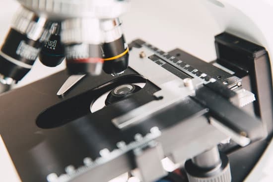
What microscope is used to see single atoms? An electron microscope can be used to magnify things over 500,000 times, enough to see lots of details inside cells. There are several types of electron microscope. A transmission electron microscope can be used to see nanoparticles and atoms.
Which type of microscope do you use to see a single atom? “So we can regularly see single atoms and atomic columns.” That’s because electron microscopes use a beam of electrons rather than photons, as you’d find in a regular light microscope.
Can you see a single atom with a microscope? Atoms are really small. So small, in fact, that it’s impossible to see one with the naked eye, even with the most powerful of microscopes. … Now, a photograph shows a single atom floating in an electric field, and it’s large enough to see without any kind of microscope. 🔬 Science is badass.
What are the two types of microscopes used to see atoms? There are two main types of electron microscope – the transmission EM (TEM) and the scanning EM (SEM). The transmission electron microscope is used to view thin specimens (tissue sections, molecules, etc) through which electrons can pass generating a projection image.
What microscope is used to see single atoms? – Related Questions
How to calculate cell size microscope formula?
L, where D is the diameter of the viewing field, E is the estimated number of cells, and L is the length of the cell.
How do objects appear microscope?
A microscope is an instrument that can be used to observe small objects, even cells. The image of an object is magnified through at least one lens in the microscope. This lens bends light toward the eye and makes an object appear larger than it actually is.
Who discovered microscope and telescope?
1590 — earliest date of a claimed Hans Martens/Zacharias Janssen invention of the compound microscope (claim made in 1655). After 1609 — Galileo Galilei is described as being able to close focus his telescope to view small objects close up and/or looking through the wrong end in reverse to magnify small objects.
What is the depth of field in a microscope?
(Science: microscopy) The depth or thickness of the object space that is simultaneously in acceptable focus. The distance between the closest and farthest objects in focus within a scene as viewed by a lens at a particular focus and with given settings.
How to focus a microscope on high power?
When focusing on a slide, ALWAYS start with either the 4X or 10X objective. Once you have the object in focus, then switch to the next higher power objective. Re-focus on the image and then switch to the next highest power.
Why is the microscope wrapped in acoustic blankets?
Why is the microscope wrapped in acoustic blankets? absorb & reflect sound. 17. What do protons determine about an element?
What is a cm microscope?
The Confocal Microscope (CM) is a simple yet considerably powerful variation of its predecessor, the conventional light microscope. In recent years the Confocal Laser Scanning Microscope has become widely established as a useful research instrument.
When would you use dark field microscope?
A dark field microscope is ideal for viewing objects that are unstained, transparent and absorb little or no light. These specimens often have similar refractive indices as their surroundings, making them hard to distinguish with other illumination techniques.
How to handle the microscope properly the do& 39?
Hold the microscope with one hand around the arm of the device, and the other hand under the base. This is the most secure way to hold and walk with the microscope. Avoid touching the lenses of the microscope. The oil and dirt on your fingers can scratch the glass.
Are cells microscopic?
Cells are so small that you need a microscope to examine them. … For most cells, this passage of all materials in and out of the cell must occur through the plasma membrane (see diagram above). Each internal region of the cell has to be served by part of the cell surface.
Who examined cork under a microscope?
As you can see, the cork was made up of many tiny units, which Hooke called cells. Cork Cells. This is what Robert Hooke saw when he looked at a thin slice of cork under his microscope.
Are atoms possible to see under a microscope?
Atoms are really small. So small, in fact, that it’s impossible to see one with the naked eye, even with the most powerful of microscopes. … Now, a photograph shows a single atom floating in an electric field, and it’s large enough to see without any kind of microscope.
What does a base do on a microscope?
Base: The bottom of the microscope, used for support Illuminator: A steady light source (110 volts) used in place of a mirror.
What does the fine adjustment do on a microscope?
FINE ADJUSTMENT KNOB — A slow but precise control used to fine focus the image when viewing at the higher magnifications.
Is bacterial pathogens too small for light microscope?
In order to see bacteria, you will need to view them under the magnification of a microscopes as bacteria are too small to be observed by the naked eye. … Bacteria have colour only when they are present in a colony, single bacteria are transparent in appearance.
What are frosted microscope slides used for?
Frosting. A procedure known as ‘frosting’ is used to elevate bond strength and ensure the most uniform thickness. Frosting maintains uniformity across the full glass slide by ensuring that the surface is parallel to the diamond grinding wheel, and creating microgrooves on the bonding side of the glass surface.
What are bright field microscopes used for?
It receives light from the light source and is responsible for the concentration of light rays on the object. Bright field microscopy is used to view fixed specimens or live cells.
When to use coarse adjustment on microscope?
COARSE ADJUSTMENT KNOB — A rapid control which allows for quick focusing by moving the objective lens or stage up and down. It is used for initial focusing.
How many microscopic organisms live on human skin?
In total, you have about 1.8 m2 of skin, and more than 1.5 trillion (that’s a 1 with 12 zeros) bacteria live on it. In some wet places, tens of millions of microbes live on every square centimetre of skin.
How microscopes work by cindy grigg?
A light (also called an optical) microscope uses a convex lens to bend light rays. A convex lens is a lens that bends outward. It is thicker in the middle than at the edges. This shape causes light rays to bend inward and meet at a point.
Can i use a magnifying glass on a microscope?
A microscope is something that uses a lens or lenses to make small objects look bigger and to show more detail. This means that a magnifying glass can count as a microscope! It also means that making your own microscope is straightforward. Microscopes use a lens or lenses to magnify objects.

