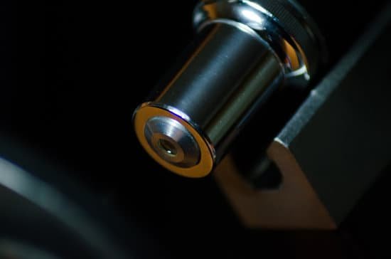
What microscope needs dead cells? The correct answer is to use a light microscope because it is the best instrument for this application. An individual can stain the cells that are specific for cell viability, for example, living cells would stain a different color than dead cells) and the resulting fluorescence can be viewed by a light microscope.
What microscope is used to see dead cells? Like the TEM, the SEM allows you to look at replicas of dead cells, after fixation and heavy metal ion staining. With this technique, electrons are reflected off the surface of the specimen.
What microscope needs a dead specimen? Comparison of the light and electron microscope
Can light microscopes see dead cells? The TEM has revealed structures in cells that are not visible with the light microscope but an electron microscope can only examine dead tissue.
What microscope needs dead cells? – Related Questions
What did robert hooke invented the first microscope?
Hooke was one of a small handful of scientists to embrace the first microscopes, improve them, and use them to discover nature’s hidden details. He designed his own light microscope, which used multiple glass lenses to light and magnify specimens. Under his microscope, Hooke examined a diverse collection of organisms.
How do you correctly use a compound light microscope?
Turn the revolving turret (2) so that the lowest power objective lens (eg. 4x) is clicked into position. Place the microscope slide on the stage (6) and fasten it with the stage clips. Look at the objective lens (3) and the stage from the side and turn the focus knob (4) so the stage moves upward.
What can a light microscope see?
Light microscopes let us look at objects as long as a millimetre (10-3 m) and as small as 0.2 micrometres (0.2 thousands of a millimetre or 2 x 10⁻⁷ m), whereas the most powerful electron microscopes allow us to see objects as small as an atom (about one ten-millionth of a millimetre or 1 angstrom or 10-10 m).
What do chloroplasts look like under a microscope?
Observation – When viewed under the microscope, students will be able to distinguish different parts of the cell including the plastids (chloroplast and mitochondria). On the other hand, a simply wet mount (even without staining) will show chloroplast to be small green (or dark green) sports across the cell surface.
What do you call a dental microscope?
A dental operating microscope (D.O.M.) has been a key tool for endodontic (root canal) dentistry since it became a requirement for residencies to teach specialists to use it in 1998.
What objective do you start with on a microscope?
When focusing on a slide, ALWAYS start with either the 4X or 10X objective. Once you have the object in focus, then switch to the next higher power objective.
Can we see dna with a microscope?
Given that DNA molecules are found inside the cells, they are too small to be seen with the naked eye. For this reason, a microscope is needed. While it is possible to see the nucleus (containing DNA) using a light microscope, DNA strands/threads can only be viewed using microscopes that allow for higher resolution.
What is working distance microscope?
■ Working Distance (W.D.) The distance between the front end of a microscope objective and the. surface of the workpiece at which the sharpest focusing is obtained.
What happens to a image in the microscope?
Microscopes invert images which makes the picture appear to be upside down. The reason this happens is that microscopes use two lenses to help magnify the image. … Images might appear to move left when you move the slide right or go up when you move them down.
How to identify amoeba under microscope?
When viewed, amoebas will appear like a colorless (transparent) jelly moving across the field very slowly as they change shape. As it changes its shape, it will be seen protruding long, finger like projections (drawn and withdrawn).
Which microscope does not allow you to view living cells?
Electron microscopes use a beam of electrons instead of beams or rays of light. Living cells cannot be observed using an electron microscope because samples are placed in a vacuum.
What does the filter do in microscope?
Microscopy filters are used to filter out specific wavelengths of light thereby increasing contrast, blocking ambient light, removing IR or UV radiation. Filters are generally fitted over the illuminating device below the iris diaphragm.
How does a binocular microscope function?
binocular microscope one with two eyepieces, permitting use of both eyes simultaneously. … compound microscope one consisting of two lens systems whereby the image formed by the system near the object is magnified by the one nearer the eye.
How do oceanic upwellings promote the growth of microscopic plankton?
How do oceanic upwellings promote the growth of microscopic plankton? They bring warmer water to the surface of the ocean. They bring dissolved nutrients to the surface of the ocean.
What is the meaning of light microscope?
The light microscope is an instrument for visualizing fine detail of an object. It does this by creating a magnified image through the use of a series of glass lenses, which first focus a beam of light onto or through an object, and convex objective lenses to enlarge the image formed.
Which microscope gives the highest amount of magnification?
Out of all types of microscopes, the electron microscope has the greatest capability in achieving high magnification and resolution levels, enabling us to look at things right down to each individual atom.
What are the parts and function of light microscope?
Lenses – form the image objective lens – gathers light from the specimen eyepiece – transmits and magnifies the image from the objective lens to your eye nosepiece – rotating mount that holds many objective lenses tube – holds the eyepiece at the proper distance from the objective lens and blocks out stray light.
What cells can we see using an electron microscope?
The cell wall, nucleus, vacuoles, mitochondria, endoplasmic reticulum, Golgi apparatus, and ribosomes are easily visible in this transmission electron micrograph.
What is an electron microscope in biology?
Electron microscopy (EM) is a technique for obtaining high resolution images of biological and non-biological specimens. It is used in biomedical research to investigate the detailed structure of tissues, cells, organelles and macromolecular complexes.
What are the steps to focus a microscope?
To focus a microscope, rotate to the lowest-power objective, and place your sample under the stage clips. Play with the magnification using the coarse adjustment knob and move your slide around until it is centered.
What cell parts can you see under a microscope?
Using a light microscope, one can view cell walls, vacuoles, cytoplasm, chloroplasts, nucleus and cell membrane. Light microscopes use lenses and light to magnify cell parts.

