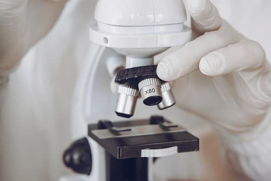
What microscope should i buy? You will need a compound microscope if you are viewing “smaller” specimens such as blood samples, bacteria, pond scum, water organisms, etc. … Typically, a compound microscope has 3-5 objective lenses that range from 4x-100x. Assuming 10x eyepieces and 100x objective, the total magnification would be 1,000 times.
What are good beginner microscopes? A scanning electron microscope would be the best choice for viewing very small surface structures of a cell.
What microscope is most useful for viewing? Lower magnification (10-20x) produces a larger field of view and is best for young kids. It is also ideal for viewing stamps and coins. Higher magnification (30-40x) is better for close-ups and more detailed work.
What are the microscopic anatomy of blood vessels? Arteries are composed of three coats: an internal or endothelial coat (tunica intima), a middle or muscular coat (tunica media), and an external or connective tissue coat (tunica adventitia). The 2 inner coats together are very easily separated from the external coat.
What microscope should i buy? – Related Questions
What is a good mp for a microscope camera?
Measurement – when measurement is the main focus we recommend selecting a camera which is not high resolution, 2 MP is usually sufficient.
How to treat microscopic hematuria?
Depending on the condition causing your hematuria, treatment might involve taking antibiotics to clear a urinary tract infection, trying a prescription medication to shrink an enlarged prostate or having shock wave therapy to break up bladder or kidney stones.
What microscope can be used to explore cells?
Two types of electron microscopy—transmission and scanning—are widely used to study cells. In principle, transmission electron microscopy is similar to the observation of stained cells with the bright-field light microscope.
What does muscle look like under a microscope?
These thick and thin filaments, when viewed under a microscope, appear “striped” or striated. This appearance under the light microscope is the reason that skeletal muscle may also be described as striated muscle. The thick and thin filaments are made up of two different contractile proteins called actin and myosin.
Who discovered microscope for the first time?
Lens Crafters Circa 1590: Invention of the Microscope. Every major field of science has benefited from the use of some form of microscope, an invention that dates back to the late 16th century and a modest Dutch eyeglass maker named Zacharias Janssen.
What is best microscope power to see hookworm eggs?
The eggs are thin-shelled, colorless and measure 60-75 µm by 35-40 µm. Figure A: Hookworm egg in an unstained wet mount, taken at 400x magnification.
Can dna be seen through a microscope?
Given that DNA molecules are found inside the cells, they are too small to be seen with the naked eye. For this reason, a microscope is needed. While it is possible to see the nucleus (containing DNA) using a light microscope, DNA strands/threads can only be viewed using microscopes that allow for higher resolution.
What is the compound light microscope most useful for viewing?
Typically, a compound microscope is used for viewing samples at high magnification (40 – 1000x), which is achieved by the combined effect of two sets of lenses: the ocular lens (in the eyepiece) and the objective lenses (close to the sample).
What microscope can be used to view living samples?
Electron microscopes are very powerful tools for visualising biological samples. They enable scientists to view cells, tissues and small organisms in very great detail.
What does many mucus in urine microscopic?
A small amount of mucus in your urine is normal. An excess amount may indicate a urinary tract infection (UTI) or other medical condition. A test called urinalysis can detect whether there is too much mucus in your urine.
What lens microscope would you see bacterial morphology?
Microscopes are optical instruments that permit us to view the microbial world. Lenses produce the magnified images that allow us to visualize the form and structure of these tiniest of living beings.
How to view live copepods under a microscope?
To observe copepods under a compound microscope, take your pipette and suck in a small amount of water from your copepod sample and place a small droplet on the slide. You should be able to see a few copepods because typically they are large enough to be seen with the naked eye.
Which paper is use to clean a microscope lens?
Another good cleaning tissue is Kodak Lens Tissue (available at photo stores) In lieu of a brush, you can use the paper. Roll the tissue into a tube and tear it in half, with the feathery torn ends together. Use it as a one-time brush. Use several for very dirty lenses.
What properties can an electron microscope see?
The shape and size of the particles making up the object; direct relation between these structures and materials properties (ductility, strength, reactivity…etc.)
How does a tunneling electron microscope work?
How an STM Works. The scanning tunneling microscope (STM) works by scanning a very sharp metal wire tip over a surface. By bringing the tip very close to the surface, and by applying an electrical voltage to the tip or sample, we can image the surface at an extremely small scale – down to resolving individual atoms.
How to calculate cell size microscope example?
Estimate how many cells laid end to end it would take to equal the diameter of the field of view. Then, divide 1,400 microns by this number to obtain an estimate of the cell’s size in microns. For example, suppose it takes 8 paramecia laid end to end to equal the diameter of the field of view.
Why might new inventions such as the telescope and microscope?
Why might new inventions such as the telescope and microscope change the way people saw the world? The telescope and microscope may have changed the way people saw the world because they allowed people to see the world in different ways.
What causes collagenous microscopic colitis?
Researchers believe that the causes may include: Medications that can irritate the lining of the colon. Bacteria that produce toxins that irritate the lining of the colon. Viruses that trigger inflammation.

