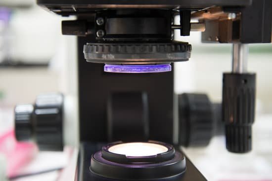
What microscope to use to see intracellular detail? Electron microscopes use a beam of electrons, opposed to visible light, for magnification. Electron microscopes allow for higher magnification in comparison to a light microscope thus, allowing for visualization of cell internal structures.
What microscope is used for internal view? A transmission electron microscope is an instrument used to create high magnification images of the internal structure of a sample being studied.
Which type of microscopy would provide the best view of these intracellular structures? Expansion microscopy (ExM) is a powerful tool which allows to visualize intracellular structures with increased resolution due to physical widening of the sample.
Which microscope will give you better detail and allow you to see inside cells? With light microscopes we can see things such as cells, parasites and some bacteria. To see much smaller things, including viruses and structures inside cells, such as DNA, we need a more powerful type of microscope. Electron microscopes use subatomic particles called electrons to magnify objects.
What microscope to use to see intracellular detail? – Related Questions
How does digital microscope work?
How does a digital microscope work? A digital microscope uses optics and a digital camera to output captured images to a computer monitor. … For instance, some of this software comes with features to record video, adjust images, edit video footage, analyze 3D samples, make measurements, and create reports.
How to look into a microscope?
Look at the objective lens (3) and the stage from the side and turn the focus knob (4) so the stage moves upward. Move it up as far as it will go without letting the objective touch the coverslip. Look through the eyepiece (1) and move the focus knob until the image comes into focus.
What is a biological microscope?
A biological microscope is generally a type of optical microscope that is primarily designed to observe cells, tissues, and other biological specimens. Multiple objective lenses can be attached, which gives these microscopes a magnification that can range anywhere from 10x – 1,000x or more.
Can microscopic mites get in lungs?
Pulmonary acariasis is a non-specific infestation of human lungs by free-living mites. In the 1930s, mites were observed in human sputum [4]. Subsequent experiments demonstrated that free-living mites can invade animal lungs and live in the respiratory tract.
What is the purpose of the arm on a microscope?
Arm connects to the base and supports the microscope head. It is also used to carry the microscope.
How is the total magnification of a compound microscope calculated?
The total magnification is calculated by multiplying the magnification of the ocular lens by the magnification of the objective lens. … Compound microscopes usually include exchangeable objective lenses with different magnifications (e.g 4x, 10x, 40x and 60x), mounted on a turret, to adjust the magnification.
How do you clean a microscope objective lens?
Place the objective lens on a dust-free surface. 2. Gently blow away loose dust that is on the surface of the optical glass with a dust blower, as if any dust left on throughout the cleaning process could scratch the optical glass or coating. Blow the air across the lens surface to avoid damaging it.
What are objectives for on a microscope?
Objectives are responsible for primary image formation and play a central role in establishing the quality of images that the microscope is capable of producing. Furthermore, the magnification of a particular specimen and the resolution under which fine specimen detail also heavily depends on microscope objectives.
How much is a tem microscope?
The cost of a transmission electron microscope (TEM) can range from $300,000 to $10,000,000. The cost of a focused ion beam electron microscope (FIB) can range from $500,000 to $4,000,000. There can be a high degree of variation in the cost of an electron microscope between manufacturers and models.
What is objective lenses of your microscope?
The objective lens of a microscope is the one at the bottom near the sample. At its simplest, it is a very high-powered magnifying glass, with very short focal length. This is brought very close to the specimen being examined so that the light from the specimen comes to a focus inside the microscope tube.
What is the pointer used for on a microscope?
Pointer: A piece of high tensile wire that sits in the eyepiece and enables a viewer to point at a specific area of a specimen.
What is a microscope slide scanner?
Microscope Slide Scanners, also referred to as Digital Pathology/Digital Histology produce quick, reliable and high-resolution images of cells. … Digital Pathology systems may provide automated cellular imaging for both fixed or live-cell assays and have modes for fluorescence, phase-contrast and transmitted light.
What cell structures can be seen with electron microscope?
The cell wall, nucleus, vacuoles, mitochondria, endoplasmic reticulum, Golgi apparatus, and ribosomes are easily visible in this transmission electron micrograph.
Can light microscopes see living things?
Light microscopes are advantageous for viewing living organisms, but since individual cells are generally transparent, their components are not distinguishable unless they are colored with special stains. Staining, however, usually kills the cells.
Who created the first microscope?
The development of the microscope allowed scientists to make new insights into the body and disease. It’s not clear who invented the first microscope, but the Dutch spectacle maker Zacharias Janssen (b. 1585) is credited with making one of the earliest compound microscopes (ones that used two lenses) around 1600.
What is the structure of microscope?
A general biological microscope mainly consists of an objective lens, ocular lens, lens tube, stage, and reflector. An object placed on the stage is magnified through the objective lens. When the target is focused, a magnified image can be observed through the ocular lens.
How big are electron microscopes?
Light microscopes let us look at objects as long as a millimetre (10-3 m) and as small as 0.2 micrometres (0.2 thousands of a millimetre or 2 x 10⁻⁷ m), whereas the most powerful electron microscopes allow us to see objects as small as an atom (about one ten-millionth of a millimetre or 1 angstrom or 10-10 m).
Who found transmission electron microscope?
Ernst Ruska at the University of Berlin, along with Max Knoll, combined these characteristics and built the first transmission electron microscope (TEM) in 1931, for which Ruska was awarded the Nobel Prize for Physics in 1986.
What type of microscope is most used in science classes?
Compound light microscopes are one of the most familiar of the different types of microscopes as they are most often found in science and biology classrooms. For this reason, simple models are readily available and are inexpensive.
What can electron microscopes see?
An electron microscope is a microscope that uses a beam of accelerated electrons as a source of illumination. … Electron microscopes are used to investigate the ultrastructure of a wide range of biological and inorganic specimens including microorganisms, cells, large molecules, biopsy samples, metals, and crystals.
What is eyepiece in microscope?
The eyepiece, located closest to the eye or sensor, projects and magnifies this real image and yields a virtual image of the object. Eyepieces typically produce an additional 10X magnification, but this can vary from 1X – 30X. Figure 1 illustrates the components of a compound microscope.

