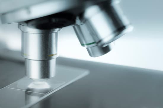
What other microscope can be used to see cell? Electrons have much a shorter wavelength than visible light, and this allows electron microscopes to produce higher-resolution images than standard light microscopes. Electron microscopes can be used to examine not just whole cells, but also the subcellular structures and compartments within them.
What microscope is used to see a cell? Two types of electron microscopy—transmission and scanning—are widely used to study cells. In principle, transmission electron microscopy is similar to the observation of stained cells with the bright-field light microscope.
Which type of microscope can be used to view cellular organelles? The electron microscope is necessary to see smaller organelles like ribosomes, macromolecular assemblies, and macromolecules. With light microscopy, one cannot visualize directly structures such as cell membranes, ribosomes, filaments, and small granules and vesicles.
What are 4 types of microscopes? There are several different types of microscopes used in light microscopy, and the four most popular types are Compound, Stereo, Digital and the Pocket or handheld microscopes. Some types are best suited for biological applications, where others are best for classroom or personal hobby use.
What other microscope can be used to see cell? – Related Questions
Who first looked at illness under a microscope?
Robert Hooke was the first to use a microscope to observe living things. Hooke’s 1665 book, Micrographia, contained descriptions of plant cells.
Can you walk after microscopic torn ligaments of the knee?
It may take 4-5 months for full healing. The patient should be able to bear weight on the knee while standing or walking, immediately after surgery. Crutches will be necessary for 2-7 days after surgery.
Which lens on a microscope makes the primary image?
The ocular lens, or eyepiece lens, is the one that you look through at the top of the microscope. The purpose of the ocular lens is to provide a re-magnified image for you to see when light enters through the objective lens. The ocular lens is generally 10- or 15-times magnification.
What can be seen with a microscope?
A microscope is an instrument that is used to magnify small objects. Some microscopes can even be used to observe an object at the cellular level, allowing scientists to see the shape of a cell, its nucleus, mitochondria, and other organelles.
Can i see sperm in a microscope?
A semen microscope or sperm microscope is used to identify and count sperm. You can view sperm at 400x magnification. … You do NOT want a microscope that advertises anything above 1000x, it is just empty magnification and is unnecessary.
What is the name of the slide on a microscope?
A glass slide is a thin, flat, rectangular piece of glass that is used as a platform for microscopic specimen observation. A typical glass slide usually measures 25 mm wide by 75 mm, or 1 inch by 3 inches long, and is designed to fit under the stage clips on a microscope stage.
Can you see cancer in a microscope?
And, unlike normal cells, cancer cells can metastasize (spread through blood vessels or lymph vessels) to distant parts of the body, too. Knowing this helps doctors recognize cancers under a microscope, because finding cells where they don’t belong is a useful clue that they might be cancer.
When was invented the first microscope?
Lens Crafters Circa 1590: Invention of the Microscope. Every major field of science has benefited from the use of some form of microscope, an invention that dates back to the late 16th century and a modest Dutch eyeglass maker named Zacharias Janssen.
Why use upright microscope?
The upright microscope has you looking down at the specimen from above. The light source and staging elements are placed beneath the slide specimen. These are great for taking a peek at fixed subjects, but they also have their limitations.
How are samples prepared for a transmission electron microscope?
For TEM, samples must be cut into very thin cross-sections. … After being fixed and dehydrated, samples are embedded in hard resin to make them easier to cut. Then, an instrument called an ultramicrotome cuts the samples into ultra-thin slices (100 nm or thinner).
Why use a scanning electron microscope?
Introduction. A scanning electron microscope uses a finely focused beam of electrons to reveal the detailed surface characteristics of a specimen and provide information relating to its three-dimensional structure. It also has a particular advantage of providing great depth of field.
What is microscopic colitis disease?
Definition & Facts. Microscopic colitis is a chronic inflammatory bowel disease (IBD) in which abnormal reactions of the immune system cause inflammation of the inner lining of your colon. Anyone can develop microscopic colitis, but the disease is more common in older adults and in women.
What is monocular and binocular microscope?
Monocular microscopes, microscopes that are equipped with one eye piece, can magnify samples up to 1,000 times. If you need a microscope that magnifies at higher levels, a binocular microscope is right for you. … Binocular microscopes have two eye pieces, which can make it easier for the viewer to observe slide samples.
What is a scanning transmission electron microscope used for?
Scanning transmission electron microscopes are used to characterize the nanoscale, and atomic scale structure of specimens, providing important insights into the properties and behaviour of materials and biological cells.
What does the body of the microscope do?
The microscope body tube separates the objective and the eyepiece and assures continuous alignment of the optics. It is a standardized length, anthropometrically related to the distance between the height of a bench or tabletop (on which the microscope stands) and the position of the seated observer’s…
Why do chloroplasts move when viewed in a microscope?
Chloroplasts migrate in response to different light intensities. Under weak light, chloroplasts gather at an illuminated area to maximize light absorption and photosynthesis rates (the accumulation response). In contrast, chloroplasts escape from strong light to avoid photodamage (the avoidance response).
What is coarse adjustment on a microscope do?
COARSE ADJUSTMENT KNOB — A rapid control which allows for quick focusing by moving the objective lens or stage up and down. It is used for initial focusing.
How does a transmission electron microscope work?
How does TEM work? An electron source at the top of the microscope emits electrons that travel through a vacuum in the column of the microscope. Electromagnetic lenses are used to focus the electrons into a very thin beam and this is then directed through the specimen of interest.
What does electron microscope consist of?
transmission electron microscope (TEM), type of electron microscope that has three essential systems: (1) an electron gun, which produces the electron beam, and the condenser system, which focuses the beam onto the object, (2) the image-producing system, consisting of the objective lens, movable specimen stage, and …
How to reuse microscope slides?
Wash slide in warm soapy water. Rinse well in running water. Blot dry on low-lint tissue (Kimwipes®) Dip into methylated spirit.
How to calculate what you can see with a microscope?
You will have to multiply the eyepiece magnification by the objective magnification to find the total magnification before dividing the field number. For instance, if your eyepiece reads 10X/22, and the magnification of your objective lens is 40.

