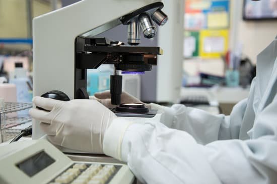
What part of the microscope moves the slide? Coarse Adjustment Knob- The coarse adjustment knob located on the arm of the microscope moves the stage up and down to bring the specimen into focus. The gearing mechanism of the adjustment produces a large vertical movement of the stage with only a partial revolution of the knob.
What part of the microscope moves the slide on the stage? Stage clips hold the slides in place. If your microscope has a mechanical stage, you will be able to move the slide around by turning two knobs. One moves it left and right, the other moves it up and down. Usually you will find 3 or 4 objective lenses on a microscope.
What part of the microscope moves the slide on the stage quizlet? Stage clips hold the slides in place. If your microscope has a mechanical stage, you will be able to move the slide around by turning two knobs. One moves it left and right, the other moves it up and down. They almost always consist of 4X, 10X, 40X and 100X powers.
Which part of the microscope moves the slide back and forth? Stage clips hold the slides in place. If your microscope has a mechanical stage, the slide is controlled by turning two knobs instead of having to move it manually. One knob moves the slide left and right, the other moves it forward and backward.
What part of the microscope moves the slide? – Related Questions
What are digital microscopes used for?
A digital microscope is an efficient tool to inspect and analyze various objects from micro-fabricated parts to large electronic devices. Digital microscopes are used in a wide range of industries, such as education, research, medicine, forensics, and industrial manufacturing.
How to calculate the field of view on a microscope?
For instance, if your eyepiece reads 10X/22, and the magnification of your objective lens is 40. First, multiply 10 and 40 to get 400. Then divide 22 by 400 to get a FOV diameter of 0.055 millimeters.
What is the purpose of the stage on a microscope?
All microscopes are designed to include a stage where the specimen (usually mounted onto a glass slide) is placed for observation. Stages are often equipped with a mechanical device that holds the specimen slide in place and can smoothly translate the slide back and forth as well as from side to side.
Where does the image get magnified in a microscope?
A microscope is an instrument that can be used to observe small objects, even cells. The image of an object is magnified through at least one lens in the microscope. This lens bends light toward the eye and makes an object appear larger than it actually is.
What is scanning tunneling microscope how does it work?
The scanning tunneling microscope (STM) works by scanning a very sharp metal wire tip over a surface. By bringing the tip very close to the surface, and by applying an electrical voltage to the tip or sample, we can image the surface at an extremely small scale – down to resolving individual atoms.
What is the purpose of a dissecting microscope?
A dissecting microscope, also known as a stereo microscope, is used to perform dissection of a specimen or sample. It simply gives the person doing the dissection a magnified, 3-dimensional view of the specimen or sample so more fine details can be visualized.
What is numerical aperture in microscope?
Numerical Aperture and Resolution. The numerical aperture of a microscope objective is the measure of its ability to gather light and to resolve fine specimen detail while working at a fixed object (or specimen) distance.
What is the resolution of transmission electron microscope?
Transmission Electron Microscope Resolution: In a TEM, a monochromatic beam of electrons is accelerated through a potential of 40 to 100 kilovolts (kV) and passed through a strong magnetic field that acts as a lens. The resolution of a TEM is about 0.2 nanometers (nm).
Why is a microscope important to epidemiology?
Microscopes form the foundation of a forensic epidemiological study. Much of the work that forensic epidemiologists conduct relates to tracking the presence of bacteria, microbes, and other molecules, organic or not within samples. Often the best tool for this job is a microscope.
Why is it desirable that microscope objectives be parfocal quizlet?
Why is it desirable that microscope objective be parfocal? So that you do not have to refocus the microscope every time you switch lenses. Which objective focuses closest to the slide when it is in focus?
What microscope can see e coli?
The nucleoid of living and OsO4- or glutaraldehyde-fixed cells of Escherichia coli strains was studied with a phase-contrast microscope, a confocal scanning light microscope, and an electron microscope.
Are bed bug egg on microscope?
Although adult bed bugs are very small, the bed bug larvae are even smaller. They appear like tiny grains of pepper and you can only see the eggs or other parts of their body by looking at them under a microscope. … In each case, they are very small and look like tinier version of their adult selves.
Which microscope uses the highest magnification?
Out of all types of microscopes, the electron microscope has the greatest capability in achieving high magnification and resolution levels, enabling us to look at things right down to each individual atom.
What part of a microscope regulates the amount of light?
Iris diaphragm dial: Dial attached to the condenser that regulates the amount of light passing through the condenser. The iris diaphragm permits the best possible contrast when viewing the specimen.
Why electron microscopes over light optical microscopes?
Electron microscopes have certain advantages over optical microscopes: Resolution: The biggest advantage is that they have a higher resolution and are therefore also able of a higher magnification (up to 2 million times). Light microscopes can show a useful magnification only up to 1000-2000 times.
What do modern scientists use microscopes for?
A microscope is an instrument that is used to magnify small objects. Some microscopes can even be used to observe an object at the cellular level, allowing scientists to see the shape of a cell, its nucleus, mitochondria, and other organelles.
Who invented microscope first time?
Lens Crafters Circa 1590: Invention of the Microscope. Every major field of science has benefited from the use of some form of microscope, an invention that dates back to the late 16th century and a modest Dutch eyeglass maker named Zacharias Janssen.
Which microscope provides the greatest resolution?
The microscope that can achieve the highest magnification and greatest resolution is the electron microscope, which is an optical instrument that is designed to enable us to see microscopic details down to the atomic scale (check also atom microscopy).
Who invented the modern compound microscope?
A Dutch father-son team named Hans and Zacharias Janssen invented the first so-called compound microscope in the late 16th century when they discovered that, if they put a lens at the top and bottom of a tube and looked through it, objects on the other end became magnified.
How is the beam focused in a scanning electron microscope?
A beam of electrons is produced at the top of the microscope by an electron gun. The electron beam follows a vertical path through the microscope, which is held within a vacuum. The beam travels through electromagnetic fields and lenses, which focus the beam down toward the sample.
Are electron microscopes dangerous?
Radiation safety concerns regarding electron microscopes are minimal, and if anything, X-ray radiation is only produced from the backscattered electrons impinging on the sample.

