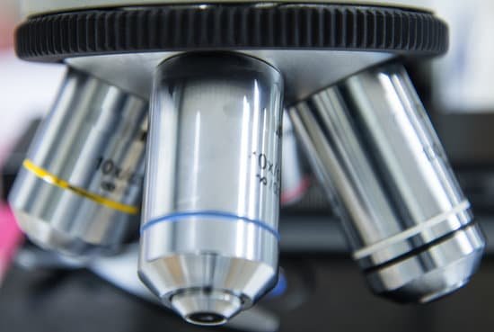
What power microscope do you need to visualize dna? To view the DNA as well as a variety of other protein molecules, an electron microscope is used. Whereas the typical light microscope is only limited to a resolution of about 0.25um, the electron microscope is capable of resolutions of about 0.2 nanometers, which makes it possible to view smaller molecules.
Can you see DNA with an optical microscope? Yes, but not in detail. “Many scientists use electron, scanning tunneling and atomic force microscopes to view individual DNA molecules,” said Michael W. … New techniques are allowing the imaging of DNA with conventional optical microscopes as well, he said, but they are in their infancy.
What can you see with 1000x microscope? At 1000x magnification you will be able to see 0.180mm, or 180 microns.
What can you see with 400x microscope? At 400x magnification you will be able to see bacteria, blood cells and protozoans swimming around. At 1000x magnification you will be able to see these same items, but you will be able to see them even closer up.
What power microscope do you need to visualize dna? – Related Questions
How to focus microscope on oil immersion?
Place a drop of immersion oil on the cover slip over that area, and very carefully swing the oil immersion lens into place. Focus carefully, preferably by observing the lens itself while bringing it as close to the cover slip as possible, then focusing by moving the lens away from the specimen.
What is the thing they put under a microscope slide?
Microscope slides are often used together with a cover slip or cover glass, a smaller and thinner sheet of glass that is placed over the specimen.
Why are microscopes important to biology?
When it comes to biology, Microscopes are important because biology mainly deals with the study of cells (and their contents), genes and all organisms. Some organisms are so small that they can only be seen by using magnifications of 40x-1000x, which can only be achieved with the use of a microscope.
What can compound light microscopes look at?
With higher levels of magnification than stereo microscopes, a compound microscope uses a compound lens to view specimens which cannot be seen at lower magnification, such as cell structures, blood, or water organisms.
What do you clean the lens of a microscope with?
Dip a lens wipe or cotton swab into distilled water and shake off any excess liquid. Then, wipe the lens using the spiral motion. This should remove all water-soluble dirt.
Is a biological cell macroscopic microscopic or submicroscopic?
Is a biological cell macroscopic, microscopic, or submicroscopic? A biological cell is microscopic, which means it is best viewed through a microscope.
Is the tem sem or compound light microscope the strongest?
The SEM has a much greater resolution capability than the light microscope because the wavelength of the electrons is about 100,000 times smaller than the wavelength of light. Resolution is the ability to distinguish between two closely-spaced points. The best resolution of the light microscope is 0.2 µm or 200 nm.
What cell parts can you see with a light microscope?
Note: The nucleus, cytoplasm, cell membrane, chloroplasts and cell wall are organelles which can be seen under a light microscope.
What is scanning tunneling microscope in biology?
The scanning tunneling microscope (STM) and the atomic force microscope (AFM) are scanning probe microscopes capable of resolving surface detail down to the atomic level. … Application of the STM for imaging biological materials directly has been hampered by the poor electron conductivity of most biological samples.
What fabric do you use to clean a microscope lens?
Once blown clean, lightly wipe the lens with Kimwipes or another approved lens cloth. Another good cleaning tissue is Kodak Lens Tissue (available at photo stores) In lieu of a brush, you can use the paper.
Can the objective lens touch the slide microscope?
Your microscope slide should be prepared with a coverslip over the sample to protect the objective lenses if they touch the slide. Do not touch the glass part of the lenses with your fingers.
Why is studying fossils with a microscope helpful?
Micropaleontologists use powerful microscopes to study fossils smaller than four millimeters (0.16 inches). Micropaleontologists often study fossils to better understand how Earth’s climate has changed. For example, they study the shells of deep-sea microorganisms.
What microscope part regulates the amount of light?
Iris diaphragm dial: Dial attached to the condenser that regulates the amount of light passing through the condenser. The iris diaphragm permits the best possible contrast when viewing the specimen.
How to calculate field of view on microscope?
For instance, if your eyepiece reads 10X/22, and the magnification of your objective lens is 40. First, multiply 10 and 40 to get 400. Then divide 22 by 400 to get a FOV diameter of 0.055 millimeters.
How to clean a microscope objective with oil contamination?
Next, soak the lens paper in a suitable solvent that can dissolve the oil and clean the lens without damaging it. We recommend anhydrous alcohol, a commercially available lens cleaning solution, or blended alcohol.
What is the lowest magnification of a light microscope?
The lowest power lens is usually 3.5 or 4x, and is used primarily for initially finding specimens. We sometimes call it the scanning lens for that reason. The most frequently used objective lens is the 10x lens, which gives a final magnification of 100x with a 10x ocular lens.
What do spherocytes look like under the microscope?
Spherocytes are round, densely staining red cells that lack central pallor and have a smaller than normal diameter. In stomatocytes, the area of central pallor is elliptical rather than round, giving the cell the appearance of the opening of a mouth (stoma).
How to view tardigrades under a microscope?
To see tardigrades under the microscope, take your wet mount, and search for them, starting with the lowest power. You should be able to see one even at 40X total magnification.
Can we see atoms with a microscope?
Atoms are really small. So small, in fact, that it’s impossible to see one with the naked eye, even with the most powerful of microscopes. … Now, a photograph shows a single atom floating in an electric field, and it’s large enough to see without any kind of microscope.
How to clean microscope lens?
Dip a lens wipe or cotton swab into distilled water and shake off any excess liquid. Then, wipe the lens using the spiral motion. This should remove all water-soluble dirt.
Can we observe nanoparticles under confocal microscope?
Confocal microscopy in combination with higher-order laser modes can detect and distinguish individual metallic nanoparticles from a single scattering image. Recently, the imaging of single metallic nanoparticles using light microscopy has attracted increasing interest.

