What rock is composed of microscopic marine algae? Diatom ooze (formed from microscopic unicellular algae having cell walls consisting of or resembling silica) is the most widespread deposit in the high southern latitudes but, unlike in the Pacific, is missing in northern latitudes.
What is Diatomite composed of? Diatomite is a soft, friable and very fine-grained siliceous sedimentary rock composed of the remains of fossilized diatoms. … Diatomite deposits formed when the skeletons of dead diatoms accumulated in either marine or freshwater environments and were subsequently compressed and lithified.
What is a marine sediment composed of? marine sediment, any deposit of insoluble material, primarily rock and soil particles, transported from land areas to the ocean by wind, ice, and rivers, as well as the remains of marine organisms, products of submarine volcanism, chemical precipitates from seawater, and materials from outer space (e.g., meteorites) …
What are the two main types based on composition of microscopic Biogenous sediments? The primary sources of microscopic biogenous sediments are unicellular algaes and protozoans (single-celled amoeba-like creatures) that secrete tests of either calcium carbonate (CaCO3) or silica (SiO2). Silica tests come from two main groups, the diatoms (algae) and the radiolarians (protozoans) (Figure 12.3.
What rock is composed of microscopic marine algae? – Related Questions
What are the parts of microscope and their uses?
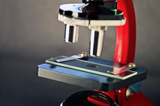
Eyepiece Lens: the lens at the top that you look through, usually 10x or 15x power. Tube: Connects the eyepiece to the objective lenses. Arm: Supports the tube and connects it to the base. Base: The bottom of the microscope, used for support.
How to measure microscope field of view?
For instance, if your eyepiece reads 10X/22, and the magnification of your objective lens is 40. First, multiply 10 and 40 to get 400. Then divide 22 by 400 to get a FOV diameter of 0.055 millimeters.
Is microscopic blood in urine bad?
This is called microscopic hematuria. Blood which is visible to the naked eye is termed gross hematuria. The chance of finding a significant urological disease is much higher in patients with gross, rather than microscopic, hematuria.
How to differentiate fungi from bacteria under microscope?
Bacteria are single-celled microscopic organisms that are characterized by the presence of incipient nucleus and few membrane-less cell organelles. Fungi, singular fungus, are eukaryotes that are characterized by the presence of chitin in the cell wall.
What are the units of cork seen under a microscope?
As you can see, the cork was made up of many tiny units, which Hooke called cells. Cork Cells. This is what Robert Hooke saw when he looked at a thin slice of cork under his microscope.
How is microscope magnification calculated?
The total magnification of the microscope is calculated from the magnifying power of the objective multiplied by the magnification of the eyepiece and, where applicable, multiplied by intermediate magnifications. … If an object is viewed with the eye from a distance of 250 mm, the magnification is 1x.
What type of microscope to detect virus?
Electron microscopy is a powerful tool in the field of microbiology. It has played a key role in the rapid diagnosis of viruses in patient samples and has contributed significantly to the clarification of virus structure and function, helping to guide the public health response to emerging viral infections.
Why are cells microscopic in size?
The important point is that the surface area to the volume ratio gets smaller as the cell gets larger. Thus, if the cell grows beyond a certain limit, not enough material will be able to cross the membrane fast enough to accommodate the increased cellular volume. … That is why cells are so small.
How do you find magnification on a microscope?
It’s very easy to figure out the magnification of your microscope. Simply multiply the magnification of the eyepiece by the magnification of the objective lens. The magnification of both microscope eyepieces and objectives is almost always engraved on the barrel (objective) or top (eyepiece).
How to connect a microscope to a computer?
Plug the device into any open USB port on the computer or the television. Hold the microscope and lightly touch the lens to the specimen. The image should now be visible on the monitor or television screen. These microscopes should only be used to examine dry specimens.
What was the first object viewed under a microscope?
The earliest microscopes were known as “flea glasses” because they were used to study small insects. A father-son duo, Zacharias and Han Jansen, created the first compound microscope in the 1590s. Anton van Leeuwenhoek created powerful lenses that could see teeming bacteria in a drop of water.
How to kill microscopic parasite trichinella?
Use a meat thermometer to ensure that the meat is thoroughly cooked. Freeze pork. Freezing pork that is less than six inches thick for three weeks will kill parasites. However, trichinella parasites in wild-animal meat are not killed by freezing, even over a long period.
What microscope would you use to examine unstained urine samples?
To obtain maximum information from the urine sample, it is important to examine the urine using phase contrast microscopy. With unstained sample, phase contrast microscopy will allow light to pass through thick or dense part of the sample which gives important information regarding the urine sample.
When was tem invented microscope?
Ernst Ruska at the University of Berlin, along with Max Knoll, combined these characteristics and built the first transmission electron microscope (TEM) in 1931, for which Ruska was awarded the Nobel Prize for Physics in 1986.
What does silk look like under a microscope?
Silk is made by the mulberry silk worm when spinning its cocoon. Under the microscope the silk fiber appears as a thin, long, smooth and lustrous cylinder. … Examined under a microscope, the cotton fibers (use a few strands of absorbent cotton) will look like a flattened, irregular, twisted ribbon.
Does ic cause microscopic hematuria?
The patient may be accustomed to an abnormal pattern. Findings on urinalysis may be entirely normal or may show microscopic hematuria or pyuria. Urine culture results are usually sterile. However, patients with interstitial cystitis may also have a concurrent bladder infection.
What brothers created the first microscope?
In the late 16th century several Dutch lens makers designed devices that magnified objects, but in 1609 Galileo Galilei perfected the first device known as a microscope. Dutch spectacle makers Zaccharias Janssen and Hans Lipperhey are noted as the first men to develop the concept of the compound microscope.
What is the difference between a microscope and a stereoscope?
A compound microscope is generally used to view very small specimens or objects that you couldn’t normally see with the naked eye. … Stereo microscopes have lower optical resolution power where the magnification typically ranges between 6x and 50x.
What do nursing informatics nurses do?
