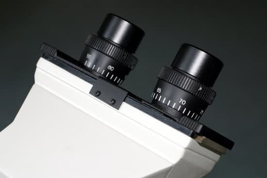
What setting should a microscope be on to see tardigrades? To see tardigrades under the microscope, take your wet mount, and search for them, starting with the lowest power. You should be able to see one even at 40X total magnification.
How can I see tardigrades? Many tardigrades are aquatic, but the easiest place for humans to find them is in damp moss, lichen, or leaf litter. Search in forests, around ponds, or even in your backyard. Your best bet is to look in damp places, where tardigrades are active.
What magnification do you need to see water? You can use a microscope with a magnification of 20x to track down water bears in a piece of moss. If you do not have a microscope at school, you can also find them with a magnifying glass with a magnification of 10x. But then the water bears will still look very small and you may not be able to see their legs.
What does a tardigrade look like under a microscope? Tardigrades are microscopic creatures that are a maximum of one millimeter in size, but usually are found to be about half that size. … They are often referred to as “Water Bears” because they look like little bears with eight puffy legs, and they have claws that look like those a grizzly bear would have.
What setting should a microscope be on to see tardigrades? – Related Questions
How does a confocal laser scanning microscope work?
A confocal microscope works with a laser and pinhole spatial filters. The laser provides the excitation light, and the laser light reflects off a mirror. … The mirrors scan the laser across the sample. The dye and the emitted light get descanned by the mirrors that scan the excitation light.
What does mucus thread mean in microscopic examination test results?
A small amount of mucus in your urine is normal. An excess amount may indicate a urinary tract infection (UTI) or other medical condition.
What can you see under a scanning electron microscope?
This technique allows you to see the surface of just about any sample, from industrial metals to geological samples to biological specimens like spores, insects, and cells.
How can you increase the resolution on a microscope?
The resolution of a specimen viewed through a microscope can be increased by changing the objective lens. The objective lenses are the lenses that protrude downward over the specimen. Grasp the nose piece. The nose piece is the platform on the microscope to which the three or four objective lenses are attached.
How does a comparison microscope work?
A comparison microscope is a device used to analyze side-by-side specimens. It consists of two microscopes connected by an optical bridge, which results in a split view window enabling two separate objects to be viewed simultaneously.
What is a compound microscope quizlet?
What is a compound microscope? -An instrument that uses light and two (or more) lenses to produce a larger image of an object.
Who invented the very first microscope?
Every major field of science has benefited from the use of some form of microscope, an invention that dates back to the late 16th century and a modest Dutch eyeglass maker named Zacharias Janssen.
What is the principle of microscope?
A simple microscope works on the principle that when a tiny object is placed within its focus, a virtual, erect and magnified image of the object is formed at the least distance of distinct vision from the eye held close to the lens.
What is revolving nosepiece in microscope?
The revolving nosepiece is the inclined, circular metal plate to which the objective lenses, usually four, are attached. The objective lenses usually provide 4x, 10x, 40x and 100x magnification. The final magnification is the product of the magnification of the ocular and objective lenses.
What is a iris diaphragm microscope function?
: an adjustable diaphragm of thin opaque plates that can be turned by a ring so as to change the diameter of a central opening usually to regulate the aperture of a lens (as in a microscope)
What are the four rules when using a microscope?
Do not touch the glass part of the lenses with your fingers. Use only special lens paper to clean the lenses. Always keep your microscope covered when not in use. Always carry a microscope with both hands.
Can you see semen under microscope?
You can view sperm at 400x magnification. You do NOT want a microscope that advertises anything above 1000x, it is just empty magnification and is unnecessary. In order to examine semen with the microscope you will need depression slides, cover slips, and a biological microscope.
How do you treat microscopic colitis?
Microscopic colitis can get better on its own, but most patients have recurrent symptoms. The main treatment for microscopic colitis is medication. In many cases, the doctor will start treatment with an antidiarrheal medication such as Pepto-Bismol® or Imodium® .
What is focusing microscope?
Focus: A means of moving the specimen closer or further away from the objective lens to render a sharp image. On some microscopes, the stage moves and on others, the tube or head of the microscope moves. Rack and pinion focusing is the most popular and durable type of focusing mechanism.
How do you determine total magnification when using a microscope?
To figure the total magnification of an image that you are viewing through the microscope is really quite simple. To get the total magnification take the power of the objective (4X, 10X, 40x) and multiply by the power of the eyepiece, usually 10X.
How many lenses did the first microscope have?
While the original microscopes that Hooke and Leeuwenhoek used may have had their limitations, their basic structure of two lenses connected by a tubes remained relevant for centuries, says Eliceiri.
What are the different types of microscope and its function?
There are several different types of microscopes used in light microscopy, and the four most popular types are Compound, Stereo, Digital and the Pocket or handheld microscopes. Some types are best suited for biological applications, where others are best for classroom or personal hobby use.
What is scanning electron microscope function?
A scanning electron microscope (SEM) scans a focused electron beam over a surface to create an image. The electrons in the beam interact with the sample, producing various signals that can be used to obtain information about the surface topography and composition.
Why do i always have microscopic blood in my urine?
Microscopic urinary bleeding is a common symptom of glomerulonephritis, an inflammation of the kidneys’ filtering system. Glomerulonephritis may be part of a systemic disease, such as diabetes, or it can occur on its own.
What is the purpose of using a microscope?
A microscope is an instrument that is used to magnify small objects. Some microscopes can even be used to observe an object at the cellular level, allowing scientists to see the shape of a cell, its nucleus, mitochondria, and other organelles.
What is the function of stage in compound microscope?
Stage is where the specimen to be viewed is placed. A mechanical stage is used when working at higher magnifications where delicate movements of the specimen slide are required. Stage Clips are used when there is no mechanical stage.

