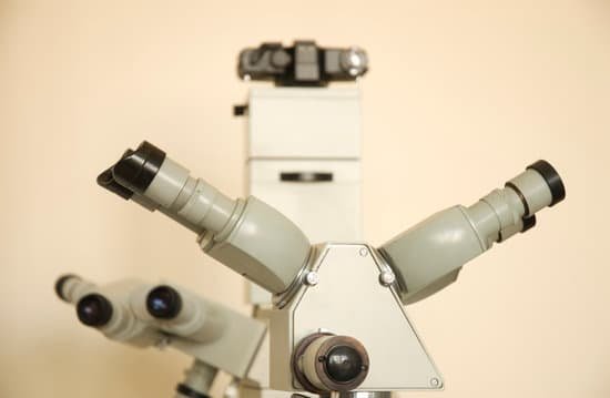
What strength microscope do you need to see sperm? You can view sperm at 400x magnification. You do NOT want a microscope that advertises anything above 1000x, it is just empty magnification and is unnecessary. In order to examine semen with the microscope you will need depression slides, cover slips, and a biological microscope.
Can you see sperm with a digital microscope? You should be able to see them, or at least see them move but they would still be very small. Also, the sample would need to be placed on a microscope slide with a cover glass and viewed on a black background. … This microscope runs up to 50X, so the sperm head would appear to be about .
Can you see sperm with a magnifying glass? The small lens can magnify 555 times, according to Science Alert, which is enough to be able to identify individual sperm cells in the video. …
What can a 1000x microscope see? A man will provide a semen sample for a semen analysis and sperm count. The doctor will check the number of sperm per sample to determine if the sperm count is too low. An ultrasound may also be performed to look for problems in the scrotum, or ducts and tubes where the semen travels.
What strength microscope do you need to see sperm? – Related Questions
How does cancer look under a microscope?
Another feature of the nucleus of a cancer cell is that after being stained with certain dyes, it looks darker when seen under a microscope. The nucleus from a cancer cell is larger and darker because it often contains too much DNA.
What is the magnification power of a dissecting microscope?
A dissecting microscope is used to view three-dimensional objects and larger specimens, with a maximum magnification of 100x. This type of microscope might be used to study external features on an object or to examine structures not easily mounted onto flat slides. Both microscopes have similar features.
Can’t see anything through microscope?
If you cannot see anything, move the slide slightly while viewing and focusing. If nothing appears, reduce the light and repeat step 4. Once in focus on low power, center the object of interest by moving the slide. Rotate the objective to the medium power and adjust the fine focus only.
How to clean a microscope eyepiece?
To clean microscope eyepiece lenses, breathe condensation onto them and then wipe them with lens tissue. Kim-wipes are made by Kleenex and generally will work well. For stubborn spots, wipe the surface with tissue moistened with 95% alcohol. Wipe the lens dry with a dry tissue.
What are the uses of a dissecting microscope?
A dissecting microscope, also known as a stereo microscope, is used to perform dissection of a specimen or sample. It simply gives the person doing the dissection a magnified, 3-dimensional view of the specimen or sample so more fine details can be visualized.
How does sperm look in a microscope?
The air-fixed, stained spermatozoa are observed under a bright-light microscope at 400x or 1000x magnification. Their viability and mor- phology can be analysed at the same time. Those appearing red-pink in colour have a damaged membrane whereas white sperm are viable, as in Photo 2.
What can you see through a light microscope?
Explanation: You can see most bacteria and some organelles like mitochondria plus the human egg. You can not see the very smallest bacteria, viruses, macromolecules, ribosomes, proteins, and of course atoms.
How much can a scanning electron microscope magnify?
An SEM can magnify a sample by about one million times (1,000,000x) at the most. Because a sample can be used in its natural state, the SEM is the easiest electron microscope to use. The final image looks 3D and shows you the outside of your sample.
What does a light microscope used for?
The light microscope is an instrument for visualizing fine detail of an object. It does this by creating a magnified image through the use of a series of glass lenses, which first focus a beam of light onto or through an object, and convex objective lenses to enlarge the image formed.
Can a microscopic exam of urine show schistosomiasis?
The gold standard for diagnosis of schistosomiasis is the microscopic detection of parasite eggs present in urine or stool1; however, parasitological diagnosis of schistosomiasis in adults is difficult, particularly among persons who have chronic infections and pass only small numbers of eggs.
Who invented the microscope in which year?
Lens Crafters Circa 1590: Invention of the Microscope. Every major field of science has benefited from the use of some form of microscope, an invention that dates back to the late 16th century and a modest Dutch eyeglass maker named Zacharias Janssen.
Who invented the first microscope to be used on cells?
In the late 16th century several Dutch lens makers designed devices that magnified objects, but in 1609 Galileo Galilei perfected the first device known as a microscope.
What can be seen using a light microscope?
Explanation: You can see most bacteria and some organelles like mitochondria plus the human egg. You can not see the very smallest bacteria, viruses, macromolecules, ribosomes, proteins, and of course atoms.
When was the microscope able to see bacteria?
Two men are credited today with the discovery of microorganisms using primitive microscopes: Robert Hooke who described the fruiting structures of molds in 1665 and Antoni van Leeuwenhoek who is credited with the discovery of bacteria in 1676.
What is the lowest possible magnification on a compound microscope?
Why do you need to start with 4x in magnification on a microscope? The 4x objective lens has the lowest power and, therefore the highest field of view.
Which parts of a microscope regulate the amount of light?
Iris Diaphragm controls the amount of light reaching the specimen. It is located above the condenser and below the stage. Most high quality microscopes include an Abbe condenser with an iris diaphragm. Combined, they control both the focus and quantity of light applied to the specimen.
What are rigid microscopes?
Features. Rigid Stands Hold Samples or Experimental Apparatuses Underneath and Around the Objective. Designed for Slides, Petri Dishes, Recording Chambers, Micromanipulators, Well Plates, and DIY Inserts. Suitable for Upright and Inverted Microscopes.
Where are the two lenses located in a compound microscope?
A compound microscope consists of two lenses, an objective lens (close to the object) and an eye lens (close to the eye).
How should a compound microscope be held and carried?
A compound microscope should be held by the arm and have one hand under the base. An image is located in the right hand corner of the FOV (field of view).
What is one disadvantage of using light microscopes?
Disadvantage: Light microscopes have low resolving power. … Electron microscopes are helpful in viewing surface details of a specimen. Disadvantage: Light microscopes can be used only in the presence of light and are costly. Electron microscopes uses short wavelength of electrons and hence have lower magnification.
How to avoid microscope touching slide?
Slide Preparation: Microscope slides should always be prepared with a cover slip or cover glass over the specimen. This will help protect the objective lenses if they touch the slide. To hold the slide on the stage fasten it with the stage clips.

