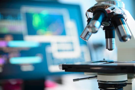
What supports the weight of the microscope? Base: This supports the weight of the microscope. Stage Clips: These hold the slide in place Stage: It’s the place where you place the slide for viewing.
Which part of the microscope supports the entire microscope? Parts of the Microscope
Which of the following parts provide balance and support the weight of a microscope? Explanation: foot is the part that provides balance and supports the weight of a microscope.
What is provides support to the microscope? The base provides stability and support for the microscope when it is upright.
What supports the weight of the microscope? – Related Questions
How to calculate total magnification with a light microscope?
Total Magnification: To figure the total magnification of an image that you are viewing through the microscope is really quite simple. To get the total magnification take the power of the objective (4X, 10X, 40x) and multiply by the power of the eyepiece, usually 10X.
Which type of microscope does not use light?
Electron microscopes differ from light microscopes in that they produce an image of a specimen by using a beam of electrons rather than a beam of light.
Which microscope has the highest resolution and magnification?
Out of all types of microscopes, the electron microscope has the greatest capability in achieving high magnification and resolution levels, enabling us to look at things right down to each individual atom.
What would cause microscopic blood in urine?
Microscopic urinary bleeding is a common symptom of glomerulonephritis, an inflammation of the kidneys’ filtering system. Glomerulonephritis may be part of a systemic disease, such as diabetes, or it can occur on its own.
What happens if you look image through a microscope?
A microscope is an instrument that magnifies an object. … The optics of a microscope’s lenses change the orientation of the image that the user sees. A specimen that is right-side up and facing right on the microscope slide will appear upside-down and facing left when viewed through a microscope, and vice versa.
How are leydig cells distinguished microscopically?
Leydig cells can be found around seminiferous tubules forming groups of up to ten cells. They are generally described as polygonal cells with eosinophilic cytoplasm and a large round nucleus with a prominent nucleolus.
When is the microscope invented?
In around 1590, Hans and Zacharias Janssen had created a microscope based on lenses in a tube [1]. No observations from these microscopes were published and it was not until Robert Hooke and Antonj van Leeuwenhoek that the microscope, as a scientific instrument, was born.
How do you get microscope coverslips clean?
Wash the coverslips extensively in distilled water (two times), then double distilled water (two times). Be sure to wash out the acid between sticked coverslips. Rinse coverslips in 100% ethanol and dry them between sheets of whatman filter paper. Keep coverslip in a clean container.
How to calculate microscopic factor?
(50 times the chamber depth of 0.02 mm) * 1000.] A variation of the direct microscopic count has been used to observe and measure growth of bacteria in natural environments.
What does fine adjustment do on a microscope?
FINE ADJUSTMENT KNOB — A slow but precise control used to fine focus the image when viewing at the higher magnifications.
What kind of microscopes used for gemology?
Gemologists usually use binocular microscopes with zoom capabilities ranging from 10 to 90 times magnification. In most circumstances, a magnification of 45x is sufficient for day-to-day operation.
What can you see in plant cells through a microscope?
They will see cell walls and chloroplasts. From the movement of chloroplasts they will infer that cyclosis, or protoplasmic streaming, is occurring. They also will observe that most chloroplasts are pressed tightly against the cell wall and should infer from this that much of the cell is occupied by a vacuole.
Why do electron microscopes have greater resolution than light microscopes?
Electron microscopes differ from light microscopes in that they produce an image of a specimen by using a beam of electrons rather than a beam of light. Electrons have much a shorter wavelength than visible light, and this allows electron microscopes to produce higher-resolution images than standard light microscopes.
Can you see amoeba without a microscope?
Most Amoeba are microscopic and cannot be seen without a microscope. However, some types of Amoeba are large enough to be seen with the unaided eye….
Can only be seen with an electron microscope?
Mitochondria are visible with the light microscope but can’t be seen in detail. Ribosomes are only visible with the electron microscope.
What is contrast on a microscope?
Contrast is defined as the difference in light intensity between the image and the adjacent background relative to the overall background intensity. …
Can you see protein under a microscope?
In a conventional optical microscope, objects less than about 200 nanometers apart cannot be distinguished from one another. … Although electron microscopes produce a detailed image of very small structures, they cannot provide an image of the proteins that make up those structures.
What is the resolution of an optical microscope?
200 nm. The good news is, there’s a difference between resolution and “ability to locate the position”. If you have one tiny and isolated fluorescent object, you can often locate the position of that object to better than your resolution.
When was the first microscope made?
Lens Crafters Circa 1590: Invention of the Microscope. Every major field of science has benefited from the use of some form of microscope, an invention that dates back to the late 16th century and a modest Dutch eyeglass maker named Zacharias Janssen.
What microscope can be used to view living specimens?
Compound microscopes are light illuminated. The image seen with this type of microscope is two dimensional. This microscope is the most commonly used. You can view individual cells, even living ones.
How do u focus a light compound microscope?
Look at the objective lens (3) and the stage from the side and turn the focus knob (4) so the stage moves upward. Move it up as far as it will go without letting the objective touch the coverslip. Look through the eyepiece (1) and move the focus knob until the image comes into focus.

