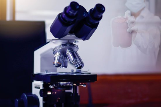
What two microscope parts affect amount of light? Iris Diaphragm is the part of the microscope that is responsible for controlling how much light reaches the ocular lens. The ocular lens is the part of the microscope through which the specimens are observed. It is also known as the eyepiece. The iris is present on the lower side of the stage.
What part of the microscope controls the amount of light? Iris diaphragm dial: Dial attached to the condenser that regulates the amount of light passing through the condenser. The iris diaphragm permits the best possible contrast when viewing the specimen.
What part changes the amount of light? Pupil: The pupil is the opening at the center of the iris through which light passes. The iris adjusts the size of the pupil to control the amount of light that enters the eye.
What are the 2 Illuminating parts of the microscope? Magnifying part – objective lens and ocular lens.
What two microscope parts affect amount of light? – Related Questions
How long does microscopic colitis last?
The outlook for people with Microscopic Colitis is generally good. Four out of five can expect to be fully recovered within three years, with some even recovering without treatment. However, for those who experience persistent or recurrent diarrhea, long term budesonide may be necessary.
When to use oil immersion in a microscope?
Oil immersion is required when viewing individual bacteria strands or details of striations in skeletal muscle. Immersion oil should be used anytime you want to view a clearer image at 1000x.
What microscope uses visible light?
The optical microscope, often referred to as the “light optical microscope,” is a type of microscope that uses visible light and a system of lenses to magnify images of small samples. Optical microscopes are the oldest design of microscope and were possibly designed in their present compound form in the 17th century.
What does the condenser do in a microscope?
On upright microscopes, the condenser is located beneath the stage and serves to gather wavefronts from the microscope light source and concentrate them into a cone of light that illuminates the specimen with uniform intensity over the entire viewfield.
How to determine the magnification when using a microscope?
To figure the total magnification of an image that you are viewing through the microscope is really quite simple. To get the total magnification take the power of the objective (4X, 10X, 40x) and multiply by the power of the eyepiece, usually 10X.
Who used microscope to see living cells?
Using a microscope that magnified objects up to about 300 times their actual size, Antony van Leeuwenhoek, in the 1670s, was able to observe a variety of different types of cells, including sperm, red blood cells, and bacteria.
Can single atoms be seen with a powerful light microscope?
Atoms are really small. So small, in fact, that it’s impossible to see one with the naked eye, even with the most powerful of microscopes.
When electron microscope invented?
Ernst Ruska, a German electrical engineer, is credited with inventing the electron microscope. The earliest electron microscope was developed in 1931, and the first commercial, mass-produced instrument became available in 1939.
Are vegetative cells green or red under microscope?
When viewed under a microscope, the endospores appear green, while the vegetative cells are red or pink. The steps in the endospore staining technique are listed below. 1. Using aseptic technique, prepare a bacterial smear on a clean slide, air dry and gently heat fix.
How does a microscope functions?
A microscope is an instrument that can be used to observe small objects, even cells. The image of an object is magnified through at least one lens in the microscope. This lens bends light toward the eye and makes an object appear larger than it actually is.
Why is a microscope used in laboratories?
The goal of any laboratory microscope is to produce clear, high-quality images, whether an optical microscope, which uses light to generate the image, a scanning or transmission electron microscope (using electrons), or a scanning probe microscope (using a probe).
What is a microspectrophotometer microscope?
The microspectrophotometer is a scientific instrument used to measure the spectra of microscopic samples. … As shown in the diagram on the left, the instrument combines a UV-visible-NIR range optical microscope with a UV-visible-NIR range spectrophotometer.
Why do microscopes flip images?
Microscopes invert images which makes the picture appear to be upside down. The reason this happens is that microscopes use two lenses to help magnify the image. Some microscopes have additional magnification settings which will turn the image right-side-up.
What is a dissecting light microscope?
A dissecting microscope is used to view three-dimensional objects and larger specimens, with a maximum magnification of 100x. This type of microscope might be used to study external features on an object or to examine structures not easily mounted onto flat slides.
What can a tem microscope see?
The transmission electron microscope is used to view thin specimens (tissue sections, molecules, etc) through which electrons can pass generating a projection image. The TEM is analogous in many ways to the conventional (compound) light microscope.
When was the first microscopic surgery?
The first microvascular surgery, using a microscope to aid in the repair of blood vessels, was described by vascular surgeon, Julius H. Jacobson II of the University of Vermont in 1960. Using an operating microscope, he performed coupling of vessels as small as 1.4 mm and coined the term microsurgery.
How to calculate magnification of microscope physics?
To calculate the total magnification of the compound light microscope multiply the magnification power of the ocular lens by the power of the objective lens. For instance, a 10x ocular and a 40x objective would have a 400x total magnification. The highest total magnification for a compound light microscope is 1000x.
When did robert hooke invent the microscope?
Robert Hooke’s Microscope. Robert Hook refined the design of the compound microscope around 1665 and published a book titled Micrographia which illustrated his findings using the instrument.
What is the diaphragm microscope?
Diaphragm or Iris: Many microscopes have a rotating disk under the stage. This diaphragm has different sized holes and is used to vary the intensity and size of the cone of light that is projected upward into the slide. There is no set rule regarding which setting to use for a particular power.
Do hospitals have microscopes?
Today, hospital laboratories use microscopes to identify which microbe is causing an infection so physicians can prescribe the proper antibiotic. They are also used to diagnose cancer and other diseases.
How does oil immersion improve resolution when using a microscope?
The microscope immersion oil decreases the light refraction, allowing more light to pass through your specimen to the objectives lens. Therefore, the microscope immersion oil increases the resolution and improve the image quality.

